Gram Positive
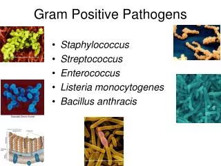
Corynebacterium Spp. Clostridium spp. Listeria spp. Bacillus spp. Gram Positive. Bacilli. Cocci. Catalase negative Streptococcaceae ( strepto - cocay -see-eye). S. viridans , S. Mutans , S. sanguis Optochin resistant Bile-insoluble No capsule. Catalase positive Staphylococcus.


Gram Positive
E N D
Presentation Transcript
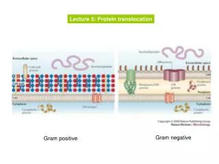
Corynebacterium Spp. Clostridium spp. Listeria spp. Bacillus spp. Gram Positive Bacilli Cocci Catalase negative Streptococcaceae (strepto-cocay-see-eye) S. viridans, S. Mutans, S. sanguis Optochin resistant Bile-insoluble No capsule Catalase positive Staphylococcus α-hemolytic (green) Coagulase positive S. Pneumoniae Optochin sensitive Bile-soluble capsule (Quellung) S. aureus Coagulase negative S. Pyogenes Group A Bacitracin sensitive β-hemolytic (clear) S. epidermidis Novobiocin sensitive γ (gamma)-hemolytic Enterococcus E. faecalis E. faecium S. agalactiae Group B Bacitracin resistant S. saprophyticus Novobiocin resistant
Methods Nasopharyngeal and oropharyngeal swabs were plated onto CNA (colistin–nalidixic acid) agar (Biolife), sheep blood agar (Biolife) and Sabouraud dextrose agar (Biolife). Sabouraud plates were incubated at 36 C in ambient air, whereas CNA and sheep blood plates were incubated at • 36 C in ambient air, • anaerobically • and in an atmosphere of 5% CO2. All plates were examined after 24, 48 and 72 h. After 24 h of incubation, α-haemolytic colonies belonging to the same streptococcal species had grown, under each of the three atmospheric growth conditions. The isolate was identified as S. pneumoniae. (Vitek2; bioMe´rieux), and confirmation of the identification was provided by inhibition of the isolate by optochin. Optochin susceptibility testing was performed in ambient air, anaerobically and in an atmosphere of 5% CO2, to prevent misidentification of Streptococcus pseudo-pneumoniaeas true S. pneumoniae (Balsalobreet al., 2006).
In Kirby-Bauer testing, white wafers containing antibiotics are placed on a plate of bacteria. Circles of poor bacterial growth surround some wafers indicating susceptibility to the antibiotic