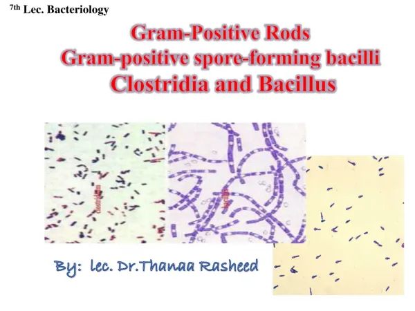
Gram-Positive Rods
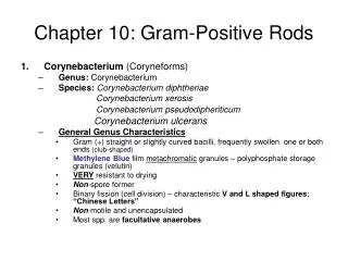
Gram-Positive Rods. Introduction. There are four medically important genera of gram-positive rods: Bacillus Clostridium Corynebacterium Listeria. Gram-Positive Rods of Medical Importance. These gram-positive rods can also be distinguished based on their appearance on Gram stain


Gram-Positive Rods
E N D
Presentation Transcript
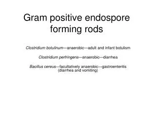
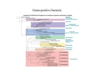
Introduction There are four medically important genera of gram-positive rods: • Bacillus • Clostridium • Corynebacterium • Listeria
These gram-positive rods can also be distinguished based on their appearance on Gram stain • Bacillus and Clostridium species are longer and more deeply staining than Corynebacterium and Listeria species • Corynebacteriumspecies are club-shaped, i.e., they are thinner on one end than the other • Corynebacteriumand Listeria species characteristically appear as V- or L-shaped rods
Non–Spore-Forming Gram-Positive Rods There are two important pathogens in this group: • Corynebacteriumdiphtheriae • Listeria monocytogenes
Disease • Diphtheria • Other Corynebacterium species (diphtheroids) - opportunistic infections
Important Properties • Gram-positive rods that appear club-shaped (wider at one end) and are arranged in palisades or in V- or l-shaped formations • The rods have a beaded appearance • The beads consist of granules of highly polymerized polyphosphate—a storage mechanism for high-energy phosphate bonds • The granules stain metachromatically, i.e., a dye that stains the rest of the cell blue will stain the granules red
Corynebacteriumdiphtheriae—Gram stain. Arrow points to a "club-shaped" gram-positive rod. Arrowhead points to typical V- or L-shaped corynebacteria
Transmission • Humans are the only natural host of Cor. diphtheriae • Both toxigenic and nontoxigenic organisms reside in the upper respiratory tract and are transmitted by airborne droplets • The organism can also infect the skin at the site of a preexisting skin lesion • This occurs primarily in the tropics but can occur worldwide in indigent persons with poor skin hygiene
Pathogenesis Exotoxin production Diphtheria toxin • inhibits protein synthesis by ADP-ribosylation of elongation factor 2 (EF-2) • The toxin affects all eukaryotic cells regardless of tissue type • The toxin is a single polypeptide with two functional domains • One domain mediates binding of the toxin to glycoprotein receptors on the cell membrane • The other domain possesses enzymatic activity that cleaves nicotinamide from nicotinamide adenine dinucleotide (NAD) and transfers the remaining ADP-ribose to EF-2, thereby inactivating it
The host response to Cor. diphtheriae consists of • A local inflammation in the throat, with a fibrinous exudate that forms the tough, adherent, gray pseudomembrane • Antibody that can neutralize exotoxin activity by blocking the interaction of fragment B with the receptors, thereby preventing entry into the cell
Schick's test • The test is performed by intradermal injection of 0.1 mL of purified standardized toxin • If the patient has no antitoxin, the toxin will cause inflammation at the site 4 to 7 days later • If no inflammation occurs, antitoxin is present and the patient is immune
Clinical Findings • The thick, gray, adherent pseudomembrane over the tonsils and throat • Nonspecific: fever, sore throat, and cervical adenopathy • Cutaneous diphtheria causes ulcerating skin lesions covered by a gray membrane. These lesions are often indolent and often do not invade surrounding tissue • Systemic symptoms rarely occur
There are three prominent complications: • Extension of the membrane into the larynx and trachea, causing airway obstruction • Myocarditis accompanied by arrhythmias and circulatory collapse • Nerve weakness or paralysis, especially of the cranial nerves
Laboratory Diagnosis • Smears of the throat swab should be stained with both Gram stain and methylene blue. The methylene blue stain is excellent for revealing the typical meta-chromatic granules • A throat swab should be cultured on Löffler's medium, a tellurite plate, and a blood agar plate • The typical gray-black color of tellurium in the colony is a telltale diagnostic criterion
An antibody-based gel diffusion precipitin test is performed to document toxin production • A PCR assay for the presence of the toxin gene in the organism can also be used
Treatment • Antitoxin-to neutralize unbound toxin in the blood • Treatment with penicillin G or an erythromycin is also recommended, but neither is a substitute for antitoxin
Prevention • Diphtheria toxoid (usually given as a combination of diphtheria toxoid, tetanus toxoid, and acellular pertussis vaccine, often abbreviated as DTaP) • Immunization consists of three doses given at 2, 4, and 6 months of age, with boosters at 1 and 6 years of age • Because immunity wanes, a booster every 10 years is recommended • Immunization does not prevent nasopharyngeal carriage of the organism