Gram Negative Rods
Gram Negative Rods. Enterobacteriaceae (Coliforms) Tests Used to ID Unknown Organisms. HOW TO ID AN UNKNOWN ORGANISM. Your goal with the enteric unknown is to determine the genus of the organism you have.

Gram Negative Rods
E N D
Presentation Transcript
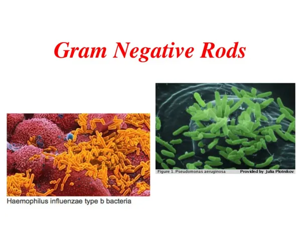
Gram Negative Rods Enterobacteriaceae (Coliforms) Tests Used to ID Unknown Organisms
HOW TO ID AN UNKNOWN ORGANISM • Your goal with the enteric unknown is to determine the genus of the organism you have. • Individual tests can be performed in separate tubes (conventional method) or all the tests can be performed in one specially designed Enterotube (rapid identification method). Comparisons between rapid identification methods and conventional methods show that they are equally accurate. • NOTE: Be able to match tests and media to their reagents, pH indicators, substrates, products, enzymes, and results, as indicated in the table at the end of the lecture transcript and flashcards.
GETTING STARTED • Perform a Gram stain. Since the longest step is air drying, make 3-4 slides and allow them to air dry at the same time, but only use one to perform your Gram stain. That way, if your culture is not decolorized properly, you have several slides ready to go so you can perform another stain quickly. When you observe your organism under the microscope, check to make sure your culture is pure. Sometimes, the Gram stain becomes contaminated or your culture may be contaminated. If you see more than one organism, you need to go back to the original pure culture and start again. If you only see one organism, the next step is to do a Negative stain with India ink, which is the best way to see the arrangements of the organism. Record the shape (rods or cocci?) and arrangement (singles, clusters, or chains?).
METHOD OVERVIEW • Determine the morphological characteristics of your unknown organism by performing a Gram stain, motility stab, capsule stain, and negative stain. If you have a Gram positive rod, you will also need to do a spore stain, since only Gram positive rods make endospores (only certain species). • Next, determine the cultural characteristics of your organism by observing the growth patterns in broth and on agar slants and plates. Determine the optimal temperature and oxygen requirements, and determine what type of hemolysis your organism displays. • Next, determine the physiological characteristics of your organism. This will require about 18-20 individual tests to find out what enzymes your organism makes, its fermentation pathway, etc.
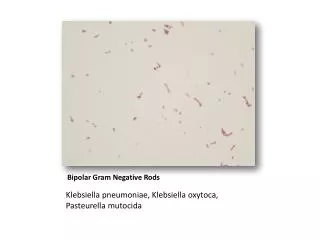
MORPHOLOGICAL CHARACTERISTICS: Gram stain, size determination, motility, capsule stain, spore stain • SIZE DETERMINATION • If you have a Gram + organism, mix a loopful of it with a loopful of a Gram neg organism whose size is known. If you have a large organism, pick a large organism to compare it with. If you have a small organism, pick a small organism. Estimate the size of yours compared to a known organism.
Controls • When performing tests on unknown organisms, control organisms are also used. • A “control” is a test media that is inoculated with a pure culture of a known organism. • One tube is inoculated with an organism that is known to be positive for that test, another tube is inoculated with an organism that is known to be negative for that test, and another tube is inoculated with the unknown organism. • When you see the results of the unknown organism, it is compared to the positive and negative control tubes to determine if the unknown is positive or negative for that test. • If the positive control or negative control did not turn the expected color, we know something is wrong with the media.
MOTILITY TEST • Inoculate a motility stab. Use a needle to obtain the inoculum. Stab the needle into the motility medium, almost all the way to the bottom, then pull the needle back out in a straight line, backing the needle out of the same stab line you made going in. Remember, these need to be incubated at room temperature (25°C). If they are placed at room temperature, the flagella will detach, giving a false negative result for motile organisms. Also remember that motility media uses TTC as a terminal electron acceptor. If the organism can use it, the media will turn red, meaning the TTC has been reduced. If there is no red color at all, you will need to do a wet mount or hanging drop to observe the organism directly to determine if it is motile. NOTE: an Enterotube can test for MRVP, citrate, indole, carbohydrate and protein catabolism, but a motility test cannot be performed in an Enterotube.
MOTILITY TEST • HANGING DROP PREPARATION • Supplies: Toothpick, petroleum jelly (Vaseline), cover slip, depression slide, gloves • Spank the bottom of the nutrient broth to mix it. • Remove one loop of broth and touch it lightly to the center of the coverslip, which is sitting on a paper towel. Try to get the smallest drop possible. • Use the toothpick to apply one small dab of Vaseline to each corner of the coverslip. This keeps the coverslip secure when it is upside down. • Clean the depression slide and press the open well down gently onto the coverslip. • Gently flip the slide over so the drop hangs in the depression well. • Since we will look under the microscope at live cells, there will be no contrast, so turn the iris down to increase the contrast. • Observe under scanning (40x); position the slide so that the edge of the drop is in the center of the field. Focus on the edge of the drop. • Increase to LP then HP, but do NOT go to 1000x because the depth of field at 1000x is too small to observe the depth of the droplet. • Look for tiny specks that are moving and record what type of motility is present.
Motility Test Positive
Motility Test • Hanging Drop Method • http://www.youtube.com/watch?v=ujzSmsmg7ok
MOTILITY TEST • Problems with hanging drop slides: • False positive results: the organism appears to move, but it cannot. This can happen if you are observing it too long under the hot light, and the liquid starts to evaporate and recede. • False negative results: the organism is capable of motility, but it does not appear to be moving. • No motility could be observed if the drop falls into the well and you don’t realize it, or if the iris diaphragm is opened too wide. Motility could be observed without false results if the culture is too concentrated.
MOTILITY TYPES • TUMBLES: the bacterium seems to be rolling over itself like a rolling stone. • RUNS: the bacterium moves from point A to point B • JIGGLES: the bacterium jiggles like it is in an earthquake, but it does not move from one part of the slide to another. THIS IS BROWNIAN MOTION, WHICH IS NOT TRUE MOTILITY. • BROWNIAN MOTION is caused by water molecules hitting a cell with low mass. It causes movement of low mass cells by the inertia created by molecular bombardment of water molecules.
NOTE: • After a person has identified if their organism is Gram positive, the next test is to do a spore stain. If it has no spores, then you need to do an Acid Fast stain to see if it is Mycobacteria. • After a person has identified if their organism is Gram negative, the next test to do is to use a thioglycollate broth to determine oxygen requirements.
CULTURAL CHARACTERISTICS: Growth patterns, temperature, hemolysis, and oxygen requirements • GROWTH PATTERNS • Use your working stock and reserve stock to observe the growth patterns of your organisms and record that information in your journal. The terminology to use is in your lab manual. When you have recorded the morphology on your reserve stock, it will be kept in the refrigerator. You will not use it except in emergency. Also use your TSB tubes from your optimum temperature experiment to determine the pattern of grown in broth.
OPTIMUM TEMPERATURE • Inoculate one loop-full of your organism into 3 TSB tubes. Label one tube 25 °C, one tube 30 °C, and one tube 38°C. Make sure your name is on the tube. These tubes will be used to determine the optimal temperature for your organism. Use the spectrophotometer to calculate their optical density at the next lab period. The tube with the most growth (highest OD) is the temperature they prefer. Organisms that grow well in room temperature as well as body temperature might be opportunistic pathogens. These tubes can also be used to determine their pattern of growth in broth.
SODIUM THIOGLYCOLATE TUBES (OXYGEN REQUIREMENT) • This medium has an oxygen gradient, which means that most of the oxygen is at the top of the tube, and the least amount of oxygen is at the bottom of the tube. To prepare this medium, a reducing agent called Sodium thioglycolate was added, which removes the free oxygen by chemically binding with it. Therefore, thioglycolate broth is called a REDUCING MEDIUM. It gets rid of the oxygen. There is also a pink indicator dye called rasazarin that shows you where the oxygen is. Notice that the pink color is only at the top of the tube. We have to be careful not to shake the tube, or we will aerate it (add more oxygen). We need the oxygen gradient to be maintained for a successful test. The results of this test determine what oxygen requirements your organism has.
SODIUM THIOGLYCOLATE TUBES (OXYGEN REQUIREMENT) • Procedure: • Hold the thioglycolate tube carefully, taking care to move it gently without shaking, jiggling, or stirring them (which introduces oxygen into the medium). • Label the tube with your name, the date, the organism, and “Thio” for Thioglycolate. • Put some of your unknown bacteria on a sterile loop and gently push the loop straight down to almost the bottom of your tube. Do not touch the bottom as this may ruin the loop, and do not introduce air by stirring or shaking the tube! • Gently pull the loop straight out of the tube and sterilize it.
SODIUM THIOGLYCOLATE TUBES (OXYGEN REQUIREMENT) • STRICT AEROBES require oxygen to grow. There will only be growth on the surface of the thio broth tube (pseudomonas and Bacillus megatarium) • STRICT ANAEROBES require the absence of all oxygen. There will only be growth at the butt (bottom) of the tube (clostridium). • FACULTATIVE ANAEROBES grow best aerobically but do not require it. Growth is throughout the tube, but is best at the top and decreases as one descends. (E.coli, staph aureus)
Thio Broth: Determines oxygen requirements This test is done to see if the organism is aerobic (growth only at the top), anaerobic (growth only at the butt), or facultative (growth throughout). A facultative organism can live with or without air, so that is considered a virulence factor.
Thio Broth Not shown: anaerobic; growth only at the butt Aerobic Facultative
CHARACTERISTICS OF ALL ENTEROBACTERACEAE (Gram neg rods in colon) • Gram negative rods • Oxidase negative • Catalase positive • Fermentation of glucose (positive) • Reduce nitrate (NO3; an inorganic substrate in the API tube. Reduction = positive) • Facultative anaerobes • If they are motile, they have peritrichous flagella, so they can run and tumble.
Amino Acids • All proteins are made of amino acids. • There are only about 22 different types of amino acids in our bodies. • Each amino acid has an amine group (NH2) and a carboxyl (acid) attached to it. Amine group: NH2 Carboxyl group: COOH “R” refers to the rest of the molecule that makes it a particular amino acid.
Amino Acids • The amino acids we will be working with in lab are these: • Tryptophan (breaks down into ammonia, pyruvic acid, and indole; causes feces smell) • Arginine (breaks down into ornithine) • Lysine (breaks down into cadaverine) • Ornithine (breaks down into putrescine) • Phenylalanine (breaks down into ammonia, pyruvic acid, and hydrogen sulfide)
Enzymes • An enzyme is a protein that cuts a substance into smaller parts. • Enzymes that cleave proteins are called protease enzymes. • Enzymes that cut an NH2 group off are called deaminases. • Enzymes that cut the carboxyl group off are called decarboxylases.
Enzymes • The substrate is the item that is cut by the enzyme. • The products are the pieces that remain after the enzyme cuts the substrate. • The pH indicator is what changes color to show us the presence of the products created by enzymes. • You need to know the substrates, products, enzymes, and pH indicators used in the tests we will discuss in lab.
PHYSIOLOGICAL TESTS FOR GRAM NEGATIVE RODS • NOTE: Know which tests have what color of a positive test: Brown, Orange or Red, Blue, Yellow, Diffused black pigment, Pink ring on top, etc. • LACTASE: this tests for an enzyme that breaks down milk sugar (lactose) into glucose and galactose. If the bacteria have this enzyme, the test is positive (yellow), but the organism is not pathogenic. Only pathogens are missing this enzyme (negative is clear). The medium is MaConkey’s agar or a lactose fermentation tube.
Lactase Test • MacConkey’s agar
Lactase Test • Fermentation Broths with Durham tubes
FERMENTATION BROTHS • If an organism has the ability to ferment sugars, the end products of the fermentation process are acids. We are looking for fermentation with acid (A) or acid + gas (AG). If there is fermentation, it will be yellow. If there is gas, the inverted miniature tube inside the media will fill with a gas bubble. If there is no fermentation, it is red, so record it as no change (NC) or Alk (protein digestion). • The medium has a Durham tube (a miniature tube that is inverted on the inside of the test tube). If gas is produced, it will form a bubble inside the inverted tube. It also has phenol red as an indicator. Phenol red turns yellow if acid is present, and red if bases are present.
FERMENTATION BROTHS • Inoculate one each of the following tubes: glucose, lactose, and sucrose. • These are different carbohydrates. After 24 hours, if the inoculated medium is yellow, it fermented the sugar in that tube. It may or may not have produced gas. Gas is produced during sugar fermentation, so when gas is present, fermentation is present as well, but not all organisms ferment with gas. If it is yellow, record it as (A). If it has gas in the Durham tube (a bubble that take up 10% of the tube, not a little bubble), record it as (AG). If it did not turn yellow (stays red), you have to look at it again in another 24 hours. After 48 hours, if the media is still red, the organism is negative for fermentation of that sugar. These tubes must be read in 24 hours, because in 48 hours, any change in color will revert to the original color.
FERMENTATION BROTHS • This is what happens: • Some organisms that ferment sugars can also digest proteins. When these organisms begin to ferment a sugar, the media becomes acidic (yellow in 24 hours), which enables them to begin digestion of the proteins which are in the media. When proteins are digested, the media becomes alkaline, and the media will turn back to red. If you want to know if it fermented the sugar, you need to read the tube in 24 hours.
FERMENTATION BROTHS • Suppose a student did not observe their tube right away, and then they see that it is red but it has gas. Since the gas is present, that indicates that it probably fermented the glucose (turned yellow at 24 hours, but he missed it), and then the organism proceeded to digest the protein, turning the media alkaline (back to red again). That would explain why it was red, but has gas (gas is produced during the fermentation process).
If Lactose Negative • Perform an Indole test (using SIM media or IMViC test): • Positive • Proteus vulgaris (H2S negative) • Providencia stuartii (H2S positive) • E. coli (Citrate negative) • Citrobacter freundii (H2S positive, Citrate positive) • Negative • Perform a urease test (all should be negative) • Perform the MR-VP test
IMViC • This stands for a series of tests: • 1) Indole (this test is also used in SIM Media) • 2) Methyl Red • 3) Voges-Proskauer • 4) Citrate • The small “i” does not stand for anything; it just makes pronunciation easier.
IMViC • The IMViC tests were developed as a means of separating members of the Enterobacteriaceae, particularly the coliforms, to determine if drinking water is contaminated with sewage. A coliform is a gram negative, aerobic or facultative anaerobic rod which produces gas from lactose within 48 hours. The presence of some coliforms indicates fecal contamination. Coliforms are only found in the GI tract of warm-blooded animals. We will perform the indole test as part of the SIM media. We performed the citrate test in the Simmon’s Citrate media. Now we need to perform the MR-VP test to complete the IMViC series.
Indole Test • The enzyme tryptophan deaminase breaks an amine group off of tryptophan (an amino acid) down into indole (which contributes to the smell of feces), pyruvic acid, and ammonia. If tryptophan deaminase is present, the indole end product reacts with Kovac’s reagent. If a red ring forms at the top of the tube, it is positive for indole, so the organism makes tryptophan deaminase. Kovac’s reagent has alcohol in it. Alcohol is lighter than water, so when the test is positive and turns red, the red ring floats to the top of the tube. (Control = E. coli). The substrate is tryptophan (an amino acid; also found in turkey). This amino acid might encourage sleep, which is why you might feel sleepy after a Thanksgiving turkey meal.
Tryptophan Deaminase (Indole) Test Tryptophan deaminase is an enzyme that slices tryptophan into indole and pyruvic acid. Add Kovac’s reagent. Red ring is positive.
MR-VP TEST (Methyl Red/Voges-Proskauer) • We do two tests with this medium: The MR test and VP test. We will inoculate one MR-VP tube today, let the culture grow until the next lab period, and then add 5 drops of Methyl Red to perform the MR test. In the next lab period, we will inoculate a new MR-VP tube, let the culture grow, and then add alpha-naphthol and potassium hydroxide reagents to perform the VP test.
MR-VP TEST • Anaerobic respiration is called fermentation. We are looking for the ability of the organism to perform glucose fermentation. Bacteria convert glucose to pyruvate using different metabolic pathways. One pathway produces unstable acidic products which quickly convert to neutral compounds. Another pathway (the butylene glycol pathway) produces neutral end products, including acetoin and 2,3-butanediol. A third pathway is the mixed acid pathway, which produces stable acidic end products which remain acidic.
MR-VP TEST • If an organism produces a lot of acid from the fermentation of sugars, it can override the buffer in the test media. If this happens, the amber media will turn red. MR-VP broth differentiates organisms that are single acid fermenters from organisms that are mixed acid fermenters because it contains over-riding buffers that affect organisms that are single acid fermenters.
MR-VP TEST • An organism that produces only one type of acid after sugar fermentation will not produce much acid, so the buffer blocks the media from changing color. But if the organism produces many different kinds of acids, it overrides the buffer and causes the color to change.
MR = METHYL RED TEST • A positive MRVP broth will be red. • Methyl Red is a yellow colored pH indicator which turns red if the organism uses the mixed acid fermentation pathway, which is that pathway that produces stable acidic end-products. If positive, the enzyme present is formic hydrogenylase. The acids will overcome the buffers in the medium and produce an acidic pH. When methyl red is added, it will go from yellow to red, which is positive for an organism that uses the mixed acid fermentation pathway.
MR Test: pH indicator Negative Positive (acid present)
MR = METHYL RED TEST • NOTE: Methyl red differs from Phenol red • Methyl Red: starts off yellow, turns redwhen acids are present (indicating glucose fermentation). Used in MR-VP test (the first part of the test) for mixed acid fermentation • Phenol Red: starts off red, turns yellowwhen acids are present (indicating glucose fermentation). Used in Urea broth and in the Fermentation broths
VP (Voges-Proskauer) TEST • The VP test is an indirect method of testing for an organism that ferments glucose using the butylene glycol pathway. Glycolysis forms pyruvic acid, which undergoes fermentation and produces acetoin, which can then be taken into one of several different fermentation pathways, depending on the organism. In the butylene glycol pathway, the end product is 2, 3 butanediol. We cannot test for that, but we can test for acetoin. If acetoin is present, it turns red (positive) and colorless if negative. A positive test means the organism uses that particular pathway for fermentation.
VP Test Negative Positive
VP (Voges-Proskauer) TEST • The VP reagents are called Barritts’s A (alpha napthol, a carcinogen!) and Barrett’s B (potassium hydroxide; KOH, a very caustic base, found in draino). If acetoin is present, it will turn a rust or red color (Gram negatives tend to do this). Therefore, red is a positive result, colorless or brown is negative.
IMViC (+ + - -)E. coli Citrate Negative Indole Positive MR Positive VP Negative
IMViC (- - + +) Enterobacter Citrate Positive Indole Negative MR Negative VP Positive