Download
1 / 23
400 likes | 1.73k Vues
GRAM POSITIVE & GRAM NEGATIVE BACTERIA. Dr . Fawzia Al-O tabi. Bacterial cells. GRAM STAIN. Developed in 1884 by the Danish physician Hans Christian Gram

E N D
GRAM POSITIVE & GRAM NEGATIVE BACTERIA Dr . Fawzia Al-O tabi
GRAM STAIN • Developed in 1884 by the Danish physician Hans Christian Gram • An important tool in bacterial taxonomy, distinguishing so-called Gram-positive bacteria, which remain coloured after the staining procedure, from Gram-negative bacteria, which do not retain dye and need to be counter-stained. • Can be applied to pure cultures of bacteria or to clinical specimens Top: Pure culture of E. coli (Gram-negative rods) Bottom: Neisseria gonorrhoeae in a smear of urethral pus (Gram-negative cocci, with pus cells)
CELL WALL Gram positive cell wall Gram negative cell wall Consists of an outer membrane containing lipopolysaccharide (LPS) thin shell of peptidoglycan periplasmic space inner membrane Lose crystal violet and stain pink from safranin counterstain • Consists of • a thick, homogenous sheath of peptidoglycan 20-80 nm thick • tightly bound acidic polysaccharides, including teichoic acid and lipoteichoic acid • cell membrane • Retain crystal violet and stain purple
Gram Positive Gram Negative
The Gram Stain Gram's Crystal iodine violet Decolorise with acetone Gram-positives appear purple Counterstain with e.g. methyl red Gram-negatives appear pink
Gram-positive rods Gram-positive cocci Gram-negative rods Gram-negative cocci
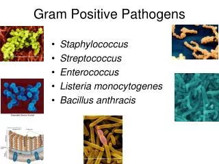
Gram positive bacteria Cocci Bacilli Aerobic /facltative Anaerobe Anaerobe Peptostreptococci Staphylococci Streptococci Enterococcci Aerobic/facultative anaerobe Anaerobic Cornybacterium Listeria Clostridium Nocardia Latobacillus ,Bacillus
Gram-positive Cocci • Staphylococci • Catalase-positive • Gram-positive cocci in clusters • Staphylococcus aureus • coagulase-positive most important • pathogen • Staph. epidermidis • and other coagulase negative staphylococci egS saprophiticus • Streptococci • Catalase-negative • Gram-positive cocci in chains or pairs • Strep. pyogenes • Strep. pneumoniae • Viridans-type streps • Enterococcus faecalis
Streptococcusdivided by type of haemolysis • S. viridans-oral flora -infective endocarditis • S. pyogenes • Group A, beta hemolytic strep • pharyngitis, cellulitis • rheumatic fever • fever • migrating polyarthritis • carditis • immunologic cross reactivity • acute glomerulonephritis • edema, hypertension, hematuria • antigen-antibody complex deposition
GRAM POSITIVE BACILLI • A-Spore forming • B-Non spore forming Spore forming are divided into:- Aerobic spore forming most important is Bacillus anthracis,that causes anthracis
Anerobic Gram Positive Bacilli • C. tetani - Tetanus C. perfringens • Gas gangarene • C. botulinum - botulism • Descending weakness-->paralysis • diplopia, dysphagia-->respiratory failure • C. diphtheriae - Fever, pharyngitis, cervical LAD • thick, gray, adherent membrane • sequelae-->airway obstruction, myocarditis
Gram-Negative Cocci • Neisseria gonorrhoeae • The Gonococcus • Neisseria meningitidis • The Meningococcus • Both Gram-negative intracellular diplococci • Moraxella catarrhalis
Gram-Negative Rods • Enteric Bacteria they ferment sugars most important are; • E. coli • Salmonella • Shigella • Yersinia and Klebsiella pneumoniae • Proteus
Gram-Negative Rods • Fastidious GNRs • Bordetella pertussis • Haemophilus influenzae • Campylobacter jejuni • Helicobacter pylori • Legionella pneumophila • Anaerobic GNRs • Bacteroides fragilis • Fusobacterium
Oxidise positive non fermentative i.e. they do not ferment sugars e.g. Pseudomonas that causes infection in Immunocompromised patients Oxidise negative non fermentative e.g. Acinobacter species
Oxidise positive comma shaped and also fermentative most important is Vibrio cholerae that causes cholera which is a disease characterized by severe diarrhea and dehydration
Non-Gram-stainable bacteria • Unusual gram-positives • Spirochaetes • Obligate intra-cellular bacteria
Unusual Gram-positives • Mycoplasmas • Smallest free-living organisms • No cell wall • M. pneumonia, M. genitalium