Download
1 / 20
341 likes | 2.46k Vues
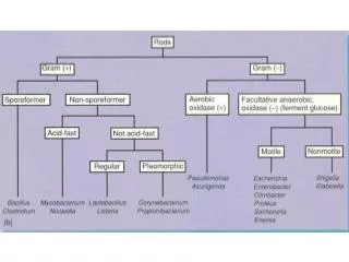
Identification Of Gram Positive Bacilli. Prepared By: Thamer Hamdan. Classification of Gram-Positive Bacilli. Gram-positive bacilli. Spore forming. Non spore forming. Aerobic. Anaerobic. Aerobic. 1- Corynebacterium 2- Listeria. Bacillus. Clostridium.

E N D
Identification Of Gram Positive Bacilli Prepared By: ThamerHamdan
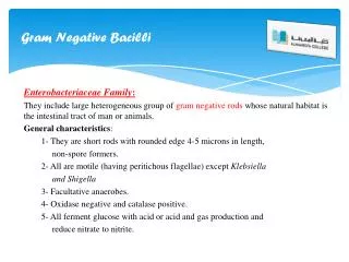
Classification of Gram-Positive Bacilli • Gram-positive bacilli • Spore forming • Non spore forming • Aerobic • Anaerobic • Aerobic • 1- Corynebacterium • 2- Listeria • Bacillus • Clostridium
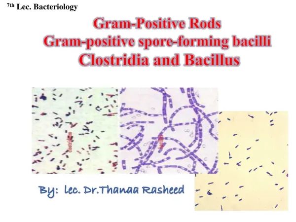
Aerobic Spore Forming Bacillus spp • Pathogenic Bacillus: -B. anthracis -B. cereus • Non-pathogenic Bacillus: - B. subtilis
General Characters of Bacillus spp • Very large Gram positive bacilli • Arranged in long chains • Motile exceptB. anthracis • Spore forming (outside the host) • Capsulated (inside the host) • Non Fastidious • Facultative anaerobic • Catalase positive • It is found in soil habitats
1- B. anthracis • B. anthracis found in soil; causes anthrax. • Transmission through inhalation of spores, breaks in skin/mucous membranes, or ingestion. • Direct person-to-person spread of anthrax is extremely unlikely to occur. 2- B. cereus • B. cereus found normally in nature. • Also isolated from food such as grains and spices • Transmission through contaminated medical equipment, or ingestion of contaminated food. • B. cereuscauses food poisoning • Many other spp. found in the environment; generally considered non-pathogens.
Identification of Bacillus Spp. • Specimens: • Fluid from cutaneous lesions • Sputum in pneumonic anthrax • Stool in intestinal anthrax (also in food poisoning by B. cereus)
Identification of Bacillus Spp. • Cultural Characteristics: • Growth on Nutrient Agar • Grow aerobically at 37 °C with characteristic mucoid or smooth colonies, which indicates the pathogensity of organism (presence of capsule). • Rough colonies are relatively avirulent. • Growth on Blood Agar • Bacillus species grow well on blood agar showing a double zone of hemolysis. • B. anthracis, which grows well on blood agar without any hemolytic effect.
Cultural Characteristics on Blood Agar: Left: B. cereus Right: B. anthracis
Differential Media for B. anthracis • PLET agar:(Polymyxin B - Lysozyme - EDTA – Thallous acetate Agar). • PLET Agar medium is the best selective medium for isolation and cultivation of B.anthracisfrom environmental specimens, animal products or clinical specimens, inhibiting B.cereus. • The colonies are circular, creamy-white with a ground glass texture.
Differential Media for B.cereus 1-Mannitol egg-yolk phenol-red polymyxin agar (MYPA): • MYPA is a selective medium for the isolation of B. cereus from faeces, vomit or food. After overnight incubation at 35-37oC, large flat, dry pinkred colonies surrounded by an area of white precipitate are produced.
2-Polymyxin egg-yolk mannitolbromothymol blue agar (PEMBA): • B. cereus produces large (5mm in diameter) and turquoise to peacock blue with a zone of egg yolk precipitation.
Identification of Bacillus Spp. • Stains: * Gram Stain • Gram positive bacilli • Found in chains • Spore is central, oval and non-bulging
* SporeStain • Bacillus spores are oval & central • By spore staining technique (Malachite green & safranin) , the spore appears green while the vegetative cells appear red.
Spore Stain Procedure:- • Make a heat fixed smear of Bacillus • Place the slide on the slide rack • Cover the smear with malachite green stain • Apply heat for 3-5 min without boiling and drying of the slide • Wash the slide gently in running water about 20 S • Counterstain with safranin for one minute • Gently rinse with water • Gently blot the slide dry, no rubbing, and let it air dry and examine with oil immersion optics. • Observe red vegetative cells and sporangia, and green endospores and free spores
Biochemical Tests: Catalase Test • All Bacillus species are catalase positive (Remember staphylococci are catalase positive)
Gelatinase test: • B. anthracis produces gelatinase in very small quantities. After inoculation and incubation of the tube which containing gelatin, the tube placed in refrigerator for approximately of 30 minutes. If the tube remains liquid that indicated positive result.
Starch Hydrolysis(Amylase Activity) • Principle • Starch + Iodineblue color • Glucose + Iodine No reaction • Nutrient Agar containing 1% Starch + M.O Glucose • Procedure • Inoculate nutrient agar plate containing 1% Starch with the M.O. • Incubate the plate at 37 for overnight • After incubation, flood the plate with Iodine solution • Result • Activity of amylase is indicated by a clear zone around the growth while the rest of the plate gives blue color after addition of iodine solution • Organism that gives (+) results is B. Subtilis & B. Cereus Amylase Iodine Appearance of colorless zone around the growth
Anaerobic Spore Forming Clostridiumspp • General Characteristics: • Gram positive, straight, thin rod with rounded ends. • All species form endospore (drumstick with a large round end). • Obligate anaerobe. • Fermentative exceptC. tetani • Motile exceptC.perfringens. • Catalase and Oxidase Negative. • Pathogenic species include: 1. C. perfringensgas gangrene & food poisoning 2. C. tetani tetanus 3. C. botulinumbotulism 4. C. difficileantibiotic associated diarrhoea