Download
1 / 83
920 likes | 1.8k Vues
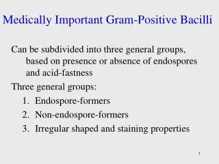
Non-Spore-Forming Gram-Positive Bacilli. Corynebacterium C. diphtheriae Disease Diphteria Opportunistic infections by other Corynebacterium species (dipheroids). Properties. Club-shaped also V- or L-shaped Beaded appearance Methachromatic granules (Albert staining) Nonmotile

E N D
Non-Spore-Forming Gram-Positive Bacilli • Corynebacterium C. diphtheriae Disease Diphteria Opportunistic infections by other Corynebacterium species (dipheroids)
Properties • Club-shaped also V- or L-shaped • Beaded appearance • Methachromatic granules (Albert staining) • Nonmotile • no capsule • Facultative anaerobic. • Classified in CNM group.
Biotypes(based on colony shape, biochemical properties and virulence) Gravis Mitis Intermedius Belfanti
Clinical finding • Common diphtheria (Nasopharyngitis) Incubation period of 2–5 days. Fibrinousexudate “pseudomembrane” Sore throat, fever, Enlargement of neck lymph nodes and neck edema. Irregulatory of cardiac rhythm, difficulties with vision, speech and swallowing. Corrosion of myelin sheaths in the central and peripheral nervous system leading to degenerating motor control
Clinical finding • Cutanous diphtheria (a secondary infection) • Antibody production: Blocking the fragment B and so preventing entry into the cell.
Transmission • Humans the only natural host • C. diphtheriae reside in the upper respiratory tract • Transmitted by airborn droplet • Infection at the site of a pre-existing skin lesion
Pathogenesis • Invasivness • Exotoxin
Invasivness • Cord factor A glycolipid inhibits eukaryotic cell oxidation. • Nuraminidase Removes N-acetyl nuraminic acid from musine membranes.
A B Exotoxin(Encoded by gen tox from a temperate phage) Fragment B. Binding of the toxin Fragment A. Enzymatic activity
Nicotinamide adenine dinucleotide phosphate (NAD) Exotoxin (A fragment) ADP Nicotinamide Reaction with EF2 Protein synthesis inhibition ADP-EF2
Testing immunity(Schick’s test) • Intradermal injection (0.1 mL): I. Cause inflammation (4-7 days later): No antitoxin in patient II. No inflammation: Antitoxin is present (Immune person)
Laboratory diagnosis • Microscopic observation (differentiation from streptococcal and vansantnasopharyngitis) • Isolating the organism Loffler’s medium a tellurite plate Tinsdal medium • Demonstrating toxin production Animal inoculation Eleck test ELISA • PCR to detect tox gene
Treatment • Tracheostomyin children (to prevent croup) • Antitoxin 20000-100000 unit (Intra muscular) • Penicillin or erythromycin
Prevention • Vaccination A combination of diphtheria toxoid, tetanus toxoid, and killed pertusis organism. Given at 2, 4 an 6 months of age, with a booster at 1 and 6 years of age and then each 10 years afterward. (DPT or DT) The toxoid is prepared by treating the exotoxin with 0.3% formaldehyde.
Listeria monocytogenes • Small rod like “chinese character” • No capsule, Facultative aerobic. • Tumbling movement. Movement in 25 c • Growing in 4c • Small and smooth colony on blood with a narrow zone of beta-hemolysis • Biochemical tests: Fermentation, Catalase + Oxidase +
Disease • Meningitis and sepsis in • The fetus or newborn as a result of transmission across the placenta or during delivery. • Immunosuppressed adults • (especially renal transplant patients) • The infected mother: asymptomatic or influenzalike illness/ Abortion
Transmission • The organism is distributed worldwide in animals, plants and soil. • Transmission to human by contact with animals or their feces unpasteurized milk contaminated vegetables. Endogenously from gasterointestinal tract.
Pathogenesis E-cadherin Phagocytosis into epithelial cells Internalin Forming filopods Phagolysosome formation (acidic condition) Inducing actin polymerization in cytoplasm Phagocytiosis By macrophages and hepatocytes LysteriolysinO secretion Release from phagolysosome
Lab. diagnosis • Microscopic observation: Diphtheroids • Isolation by culture: Blood and CSF samples on blood agar Colonies: Small, gray colonies with a narrow zone of beta hemolysis
Treatment • Penicillin Resistant are rare Prevention • Cell-mediated immunity is active but no immunization • Limiting the exposure of immunosuppressed patients to potential sources
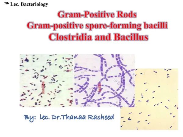
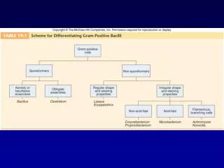
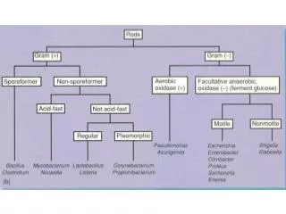
Spore-forming gram-positive bacilli • Bacillus (Aerobic) B. antheracis, B.cereus • Clostridum (Anaerobic) C. tetani, C. botulinum, C. perfringens, C. difficile
Bacillus anthracis • Disease Anthrax (common in animal but rare in humans).
Properties • A large rod with square ends. • Frequently in chains • A unique anti-phagocytic capsule is composed of D-glutamate. • Non-motile (other members of the genus are motile.)
Transmission • Spores persist in soil for years. Infection from animal products (hides, bristles and wool), contact with sick animal. • Portals of entry: skin, mucous membranes, and respiratory tract.
Clinical findings • A typical lesion: A painless ulcer with black, necrotic eschar. Local edema. • Untreated cases progress to bacteremia and death. • Woolsorter’s disease (pulmonary anthrax) is a life threatening pneumonia (by inhalation of spores).
Pathogenesis • Invasiveness • Exotoxin • Anthrax toxin, has 3 components: • Protective antigen • Lethal factor: In the presence of protective antigen is rapidly fatal for mice. The action is unknown • Edema factor (an exotoxin): An adenylatecyclase dependent on protective antigen for its binding and entry into the cell.
Lab. diagnosis • Samples: Exudate, Blood, sputum. • Direct smear: Large rods in chains. Spores not seen in smears of exudate. • Culture and biological/biochemical tests (Sensitivity to penicillin (String of pearls test), Fermentation, gelatin hydrolysis, Motility) • No serological tests are useful
Prevention • Preventing soil contamination • Sterilizing dead animals and animal products . • Protecting persons at risk of exposure with special clothes. • Vaccination with cell-free vaccine for persons at high risk.
Treatment • Penicillin No resistant strain isolated
Bacillus cereus • Motile • No capsule • Saprophyte