Download
1 / 35
390 likes | 1.26k Vues
Diagnostic microbiology lecture: 13 Gram Positive, Non Endospore-Forming Bacilli CORYNEBACTERIUM Abed ElKader Elottol MSc. Microbiology 2010. Medically Important SPECIES: 1. Corynebacterium diphtheria 2. Corynebacterium ulcerans 3. Corynebacterium ovis (pseudotuberculosis).

E N D
Diagnostic microbiologylecture: 13Gram Positive, Non Endospore-Forming Bacilli CORYNEBACTERIUM Abed ElKader Elottol MSc. Microbiology 2010 abed elkader elottol
Medically Important SPECIES: 1. Corynebacterium diphtheria 2. Corynebacterium ulcerans 3. Corynebacterium ovis (pseudotuberculosis) abed elkader elottol
Corynebacterium diphtheria • Clinical manifestation: • Local lesions in the nose and throat. • Primary infection may affect ear, conjuctiva, umbilicus and vagina. • The classic form of the disease is characterized by a local pseudomembraneous. • lesion covering the tonsils, pharynx or in the nose. • The pseudomembrane can extend into trachea and completely obstruct the air passage, causing the patient to die of suffocation. • Diphtheria toxin produced in the local lesion which is absorbed systematically can damage such distant organs and tissues such as heart, liver, kidneys and central nervous system. • Death is usually results from heart failure. abed elkader elottol
Sign and Symptoms • The respiratory form has an incubationperiod of 2-5 days. • The onset of disease is usually gradual. • Symptoms include fatigue, fever, a mild sore throat and problems swallowing. • Children infected have symptoms that include nausea, vomiting, chills, and a high fever, although some do not show symptoms until the infection has progressed further. • In 10% of cases, patients experience neck swelling, informally referred to as "bull neck." • These cases are associated with a higher risk of death.
General Characteristics: • Gram-positive bacilli, Pleomorphic (Curved or straight). • Non-motile, non-acid fast and aerobic. • Club-shaped forms = Swelled end • Chinese characters “Cell arrangement L or V shapes are also observed ). • Ability to grow on selective media containing potassium tellurite. • Specimen Collection: • Throat and nasopharengeal. • When diagnosis is confirmed, patient contacts must be subjected to the same procedures. • Moisten swabs with Normal saline and collect in the usual manner. abed elkader elottol
Media Pai Medium Composition per 1620.0mL: Glucose...............................................................5.0g Homogenized whole egg...................................1.0L Glycerol......................................................120.0mL pH 6.75 ± 0.2 at 25°C Use: For the isolation and cultivation of Corynebacterium spp. abed elkaderelottol
Tinsdale Agar Composition per 1100.0mL: peptone............................................................20.0g Agar..................................................................15.0g NaCl....................................................................5.0g Yeast extract........................................................5.0g L-Cystine...........................................................0.24g Tinsdale Supplement: Composition per 100.0mL: Na2S2O3(Sodium Thiosulphate )................... 0.43g K2TeO3(Potassium Tellurite) ....................... 0.35g Serum..........................................................100.0mL Use: For the primary isolation and identification of Corynebacterium diphtheriae. The formation of black to brown halos surrounding the colony results from the reduction of potassium tellurite to metallic tellurite. abed elkaderelottol
Loeffler Medium Composition per liter: Beef serum........................................................70.0g Egg, dried............................................................7.5g Heart muscle, solids from infusion...................0.72g Glucose.............................................................0.71g Peptic digest of animal tissue...........................0.71g NaCl..................................................................0.36g pH 7.6 ± 0.2 at 25°C Use: For the cultivation of Corynebacterium diphtheriae. For demonstration of pigment production and proteolysis by Corynebacterium diphtheriae abed elkader elottol
Hoyle Medium Base Composition per liter: Agar..................................................................15.0g Beef extract.......................................................10.0g Peptone.............................................................10.0g NaCl....................................................................5.0g Blood…………....................................................50.0mL Tellurite solution...........................................10.0mL pH 7.8 ± 0.2 at 25°C Tellurite Solution: Composition per 100.0mL: K2TeO33.5g................................................................ Caution: Potassium tellurite is toxic. Use: For the isolation and differentiation of Corynebacterium diphtheriae abed elkaderelottol
Pai medium and Loeffler medium provides good growth of C.diphtheria and stained smears prepared from both media may show the characteristic Chinese letters blue bacillus. Colony Morphology: Gray to black with irregular edges on Loeffler medium. Based on colony morphology Corynebacteria are divided into 3 groups on Tinsdale Agar and Hoyle Medium 1. Gravis: Large, flat, gray to black, non-hemolytic 2. Mitis: Smaller, more convex, darker with dull surface 3. Intermedius: The smallest of all. Note: Gravis strain hydrolysis starch and glycogen while mitis and intermedius do not. abed elkader elottol
Diphtheria toxin: • Structure: • It is a heat-labile polypeptide that can be lethal in a dose of 0.1 ug/kg. • If disulfide bond are broken, the molecule can give two fragments. (A & B). • Fragment B has no independent action but it is required for the transport of fragment A. • 2. Mechanism of Action: • Fragment A inhibits polypeptide chain elongation by inactivating the elongation factor 2 "EF2". • This factor is required for translocation of polypeptidyl-transfere RNA from the acceptor to the donor site on the eukaryotic ribosome. • Thus preventing protein synthesis leading to cell death. abed elkader elottol
Biochemical tests: The colonial morphology of C. diphtheria on Tinsdale medium is the most important characteristics in differentiating it from other species of corynebacteria. =Urease Negative =Nitrate Positive =Gelatin liquefaction: Negative Fermentation reactions of various carbohydrates. abed elkader elottol
Toxigenicity Tests: • 1. In-Vivo toxigenicity test: • 1. From a pure culture of an isolate, inoculate a 10-ml tube of BHIB, incubate at 370C for 48 hours. • 2. Inject one guinea pig with diphtheria antitoxin to serve as a control. • 3. After 2 hours, inject 5 ml of the broth into both the control and test animals. • 4.Observe after 48 hours. • Results and interpretation • If the test animal dies and the control survives= It is C. diphtheria • If both the test and control show illness or die= It is another organism. abed elkader elottol
2. In Vitro toxigenicity test: Principle: This test is based on a precipitin reaction between diphtheria toxin produced by the test culture and diphtheria antitoxin in the medium. The reaction produces insoluble products that precipitate. Procedures: 1. Paper strip is saturated with diphtheria antitoxin. 2. The medium contained in a petridish is streaked with the test organism or organisms if more than one strain are to be tested. 3. Place the paper strip perpendicular to the organism streaks. 4. Incubate the plates for 24 hours at 37 oC. 5. Examine the line of precipitation that extend out from the line of bacterial growth. 6. The presence of line of precipitation is considered positive and it confirms that the test organism as a toxigenic. abed elkader elottol
Skin test = Schick test To determine whether The patient is susceptible to diphtheria infection or not by detecting the presence or absence of antibodies. Procedures: 1. 0.1 ml of toxin is injected intradermally in one arm. 2. 0.1 ml of heated (inactivated) toxin as a control is injected in the other arm. 3. Read after 24-48 hours. Positive: (Susceptible)Redness and swelling that increases for several days and then fades, leaving brownish pigmented area. The control site shows no reaction. Negative:(Not susceptible) Neither injection site shows any reaction). abed elkader elottol
Treatment: • Antibiotic therapy: Penicillin or erythromycin To eradicate the organism. • 2. Anti-toxin: To neutralize the effect of diphtheria toxin. • Control: • By immunization with inactivated or attenuated toxin of toxigenicC.diphtheria. abed elkader elottol
Erysipelothrix insidiosa E. Rhusiopathiae Abed ElKader ElOttol
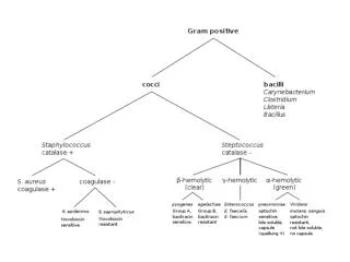
Disease: Erysipleoid (red skin) it has three distinct clinical entities: 1. Erysipeloid : Localized cutaneous infection usually on the finger and hands. 2. Generalized cutaneous infection. 3. Septicemia Microorganism: - Gram-positive rods. - Non-motile. - Non-spore forming. - Facultative anaerobe. Abed ElKader ElOttol
Specimen and media: • Usually skin biopsy from the advancing borders of the lesion. • Blood specimen if septicemia is suspected. • -Culture media: 1% glucose broth and blood agar. • NB: Make gram staining for the organism from the clinical specimen and the growth on blood agar. • Biochemical tests used in the identification: • Test tube brush appearance in gelatine stabs. • H2S production. • Acidification of slant and butt in TSIA with no gas production. • To differentiate this organism from Listeria monocytogenes, perform the catalase test. Listeria monocytogenes is catalase Positive. • Treatment: • - Penicillin is the drug of choice. Abed ElKader ElOttol
Listeria monocytogenes Abed ElKader ElOttol
Disease: Listerosis = it has four forms 1. Non-specific flu-like illness during pregnancy or puerperal sepsis. 2. Neonatal sepsis and later meningitis. 3. Sepsis or meningitis in immunocompromised patients. 4. Food poisoning. Microorganism: - Gram-positive rods (Shorter & Thinner). - Non-spore forming - Facultative anaerobe - Beta hemolytic on Blood agar. Abed ElKader ElOttol
Biochemical Characteristics: • Motile (Umbrella-type growth in semi-solid media) • Motile (Tumbling motility as seen by the hanging drop technique) • Catalase positive • V.P positive • Specimen and Culture media: • Blood, cerebrospinal fluid (CSF) and genital tract secretion. • Media : BHIA + 5% sheep blood and BHIB. • Selective Listeria Agar SLA • = Brownish to black discoloration is usually seen around the colonies of Listeria monocytogenes. Abed ElKader ElOttol
Identification: 1. The demonstration of Gram-positive bacilli from stained smears (CSF and other fluids or food samples). 2. Culture and Isolation: On Blood Agar or Selective Listeria Agar. 3. Biochemical tests: Catalase test, V.P, and motility test. 4. Serological test: Agglutination with specific sera. Treatment: Combined therapy of penicillin plus aminoglycosides. Tetracycline also may be used as a second line if the patient is sensitive to penicilllin or aminoglycosides. Abed ElKader ElOttol
The same Blood Agar plate examined with transmitted light. The colonies are surrounded by a narrow haemolytic zone. This zone is more easily seen if the culture is grown on thin Blood Agar plates. Pinpoint to small, semi-transparent colonies. abed elkader elottol
Listeria monocytogenes grows as brown-green colored colonies with a black halo (esculin splitting). abed elkader elottol
END of LECTURE abed elkader elottol