Download
1 / 28
370 likes | 1.14k Vues
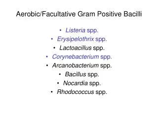
Aerobic/Facultative Gram Positive Bacilli. Listeria spp. Erysipelothrix spp. Lactoacillus spp. Corynebacterium spp. Arcanobacterium spp. Bacillus spp. Nocardia spp. Rhodococcus spp. Corynebacterium. Mycolic acids: Present in cell wall

E N D
Aerobic/Facultative Gram Positive Bacilli • Listeria spp. • Erysipelothrix spp. • Lactoacillus spp. • Corynebacterium spp. • Arcanobacterium spp. • Bacillus spp. • Nocardia spp. • Rhodococcus spp.
Corynebacterium • Mycolic acids: Present in cell wall • Metachromatic granulescanbe observed in rods stained with methylene blue • Metabolism: Fermentative • “Club”-shaped, non-motile, catalase positive • Most species grow aerobically on most media • Lipophilic strains require lipids for good growth
Corynebacterium Species • Non-Lipophilic • Corynebacterium diphtheriae • Corynebacterium amycolatum • Corynebacterium striatum • Corynebacterium pseudodiphtheriticum • Lipophilic • Corynebacterium jeikeium • Corynebacterium urealyticum
C. diphtheriae Pathogenesis and Immunity C. diphtheriae occurs in the respiratory tract, in wounds, or on the skin of infected persons or normal carriers. It is spread by droplets or by direct contact. Portal of entry: respiratory tract or skin abrasions. Diphtheria bacilli colonize and grow on mucous membranes, and start to produce toxin, which is then absorbed into the mucous membranes, and even spread by the bloodstream. Local toxigenic effects: elicit inflammatory response and necrosis of the faucial (咽喉的) mucosa cells-- formation of "pseudo-membrane“ (composed of bacteria, lymphocytes, plasma cells, fibrin, and dead cells), causing respiratory obstruction. Systemic toxigenic effects: necrosis in heart muscle, liver, kidneys and adrenals. Also produces neural damage.
Diphtheria toxin is an A-B toxin expressed from a temperate phage (b-phage) in the presence of low iron concentrations. This toxin binds to receptors on the surface of many eukaryotic cells, particularly heart and nerve cells, and results in inhibition of polypeptide chain elongation by ribosylation of the elongation factor EF-2. It can induce protective antibodies (antitoxin). Emil von Behring, Shibasaburo Kitasato : Diphtheria antitoxin serum, 1901 Noble Prize
Emil von Behring discovered that guinea pigs injected with diphtheria toxin (remnants of diphtheria with the active bacilli filtered out) can have their tissues acclimated(適應) to the toxin such that they produce a substance capable of neutralizing the diptheria toxin itself. This antitoxin, he mixed with diphtheria toxin and injected into healthy guinea pigs, yielding no ill effects after the animals were exposed to diptheria. When this treatment was applied to humans, the mortality from that disease was reduced to a negligible level.
Diphtheria Toxin: Structure • Toxin of 58,342 daltons molecular weight • Subunit B • 21,500 daltons • receptor-binding domain and translocation domain • Subunit A • catalytic domain
Diphtheria Toxin: Mode of Action • The heparin-binding epidermal growth factor on the cell surface is the receptor for the toxin • After attached to the cell, the toxin is taken up by endocytosis • Acidification of the endocytic vesicle allows unfolding of the A and B chains exposing the translocation domain (T domain) of the toxin • The T domain inserts into the endosome membrane translocating the A subunit into the cytoplasm where it regains its enzymatic configuration • The enzymatic A subunit inactivates elongation factor 2 (EF-2) and terminates host cell protein synthesis • One EF-2 molecule per ribosome in a cell, so one exotoxin molecule can completely turn off protein synthesis
C. diphtheriae Clinical Diseases Respiratory diphtheria Incubation period: 2-6 days. Inflammation begins in the respiratory tract, causing sore throat, exudative pharyngitis that develops into pseudomembrane, and low grade fever. Prostration (衰弱) and dyspnea (呼吸困難)soon follow, which may lead to suffocation if not promptly relieved by intubation or tracheotomy(氣管切開術). Damage to the heart causes irregular cardiac rhythm. Visual disturbance, difficulty in swallowing and paralysis of the arms and legs also occur but usually resolve spontaneously. Death may be due to asphyxia(窒息)or heart failure. Cutaneous diphtheria: mild (papule (丘疹 ) ulcer with grayish membrane) with little toxigenic effects. Stimulates antitoxin production.
C. diphtheriae Laboratory Diagnosis Specific treatment should be given before the lab reports if the clinical picture strongly suggests diphtheria. Specimens: swabs from the nose, throat or suspected lesions. Gram's stain: beaded rods in typical arrangement (unreliable). Culture: inoculate specimen onto a cysteine-tellurite blood agar plate. Identification: biochemical tests (presence of cysteinase). Toxigenicity test: 1. in vivo test: inject the culture into antitoxin-protected and unprotected guinea pigs subcutaneously. 2. Tissue culture neutralization assay. 3. in vitro test: immunodiffusion assay (Elek test ). 4. Detection of toxin gene by PCR.
Elek Test sterile filter paper with C. diphtheriae antitoxin Ab-Ag precipitation line bacteria
C. diphtheriae Treatment Treatment of diphtheria rests on prompt administration of antibiotics (penicillin, erythromycin) and diphtheria antitoxin. Maintenance of an open airway. Treatment of bacteremia or endocarditis must be guided by antibiotic susceptibility tests.
C. diphtheriae Prevention and Control Humans are the only known reservoir of C. diphtheriae. Diphtheria was mainly a disease of small children. This organism is maintained in the oroparynx or skin ofasymptomatic carriers. The bacteria are spread directly from person to person. To limit contact with diphtheria bacilli to a minimum, patients with diphtheria should be isolated. Prophylactic antibiotic treatment to unimmunized contacts.
C. diphtheriae Prevention and Control Active immunization in childhood with diphtheria toxoid yields antitoxin levels adequate until adulthood. Usually combined with tetanus toxoid and/or pertussis vaccine (DPT vaccine). All children must receive an initial course of immunizations and boosters (5 injections at 2 months, 4 months, 6 months, 15-18 months, and at 4-6 years). Regular booster (every 10 years) with Td (tetanus and diphtheria) toxoids are particularly important for adults who travel to developing countries.
Other Corynebacterium Species They are ubiquitous in plants and animals. Many are found as part of human normal flora and may cause opportunistic infections, such as pneumonia, endocarditis, and soft tissue and bone infections, in immunocompromised patients. C. jeikeium: sepsis, endocarditis, wound infections, foreign body infections. C. urealyticum causes UT infections. It is a strong urease producer, infection of UT may lead to formation of stones. C. ulcerans is closely related to C. diphtheriae. May cause diphtheria-like disease. Resistant to many antibiotics. Treatment of bacteremia or endocarditis must be guided by antibiotic susceptibility tests.
Listeria and Erysipelothrix L. monocytogenes: meningitis and bacteremia E. rhusiopathiae: erysipeloid Structure and Physiology of Listeria Small gram-positive coccobacilli; facultative anaerobe. Motile at room temperature but not at 37oC. Grow on most conventional media in a wide pH range and cold temperatures.
L. monocytogenes Pathogenesis and Immunity Widely distributed in nature (soil, water, vegetation, and the intestines of a variety of animals).Fecal (排泄物的)carriage in healthy people: 1%-5%. Human disease is rare and is restricted to neonates and the elderly, pregnant women, and immunocompromised patients (particularly those with defective cell-mediated immunity, such as AIDS patients). Infection may be initiated in the intestine. Facultative intracellular pathogen. The intracellular survival and spread of the bacteria are critically important in pathogenesis and, therefore, cellular immunity is more important than humoral immunity in host defense against this organism.
Internalins- interaction with host cell Listeriolysin O- release the bacteria to cytosol ActA – penetrate to adjacent cell
L. monocytogenes Clinical Diseases Adults Healthy Asymptomatic or mild influenza-like illness. Gastrointestinal symptoms in some patients. Immunocompromised Meningitis (high risk: organ transplant patients, cancer patients, pregnant women) Primary bacteremia: chills and fever; high fever and hypotension in severe cases. Maybe fatal. Neonates Early onset disease (acquired transplacentally in utero): granulomatosis infantiseptica(敗血嬰兒肉芽腫), with disseminated abscesses and granulomas in multiple organs. Late onset disease (acquired at or soon after birth): meningitis or meningoencephalitis with septicemia, similar to that caused by group B streptococci.
L. monocytogenes Laboratory Diagnosis Specimen: CSF and blood. Gram stain: CSF typically show no Listeria because of the low bacterial concentration. Culture Listeria grows on most conventional media. Selective media and cold enrichment are used for specimens contaminated with rapidly growing bacteria. Hemolysis (b-) and motility in liquid or semisolid medium are useful for preliminary identification. Identification Biochemical and serological tests.
L. monocytogenes Treatment, Prevention, and Control L. monocytogenes is resistant to multiple antibiotics (e.g., cephalosporin and tetracycline). Currently, penicillin or ampicillin, either alone or with gentamicin, is the treatment of choice. Outbreaks have been associated with the consumption of contaminated milk, soft cheese, undercooked meat, unwashed raw vegetables, and cabbage. Refrigeration of contaminated food products permits the slow multiplication of the organisms to an infectious dose. Because Listeria organisms are ubiquitous and most infections are sporadic, prevention and control are difficult. High risk people should avoid eating raw or partially cooked foods.
Food-Borne Listeriosis • Present in raw milk and vegetables, fish, poultry, fresh and processed meats and fish • Other implicated foods have included cabbage, cole slaw, soft cheeses, and shrimp
Erysipelothrix(Hair of red disease) E. rhusiopathiae Slender gram-positive, microaerophilic, with a tendency to form filaments. Form small, grayish a-hemolytic colonies after 2 to 3 days incubation. Widely distributed in wild and domestic animals. Animal disease (particularly in swine) is widely recognized, but human disease is uncommon. Causes zoonotic infections through an abrasion or wound: Localized skin infection (erysipeloid): 1-4 day incubation; painful and pruritic (瘙癢), slowly spreading inflammatory skin lesions on the fingers or hands, violaceous with raised edge. Suppuration is uncommon (distinguishing from Streptococcal erysipeals) . Generalized (diffuse) cutaneous infection: rare and often associated with systemic manifestation. Septicemia: uncommon and frequently associated with endocarditis.
Erysipelothrix Penicillin is the antibiotic of choice. Specimen: full-thickness biopsy specimens or deep aspirates (because the bacteria locate only on deep tissues). Culture: grow on most conventional media in the presence of 5%-10% CO2. Identification Motility- and catalase-negative. People at occupational risk (butchers, meat processors, farmers, poultry workers, fish handlers, and veterinarians) are prevented by use of gloves and other coverings on exposed skin. Vaccination is used to control disease in swine.