Download
1 / 52
540 likes | 1.34k Vues
Haemophilus , Legionella and Bordetella Gram Negative Rods of the Respiratory Tract . Haemophilus. Introduction. During the influenza pandemic of 1918 a “ pleomorphic ” Gram negative bacterium was isolated from the respiratory tract of most of the patients who were dying of influenza

E N D
Haemophilus, Legionella and Bordetella Gram Negative Rods of the Respiratory Tract
Introduction • During the influenza pandemic of 1918 a “pleomorphic” Gram negative bacterium was isolated from the respiratory tract of most of the patients who were dying of influenza • The bacterium grew only on media containing blood from rabbits, horses, and cows. It did not on media containing unheated sheep’s blood unless it was first heated to 80oC, a temp that releases X & V factors without destroying them. This was the 1st use of what we now call chocolate blood agar (CBA). • The bacterium was named Haemophilus, or “blood loving” because it grew only on media containing blood. The species name “influenzae” was used because it was mistakenly presumed to cause influenza. • In the 1930s, after cell cultures and the electron microscope were invented, it was demonstrated that influenza is caused by a virus, the influenza virus (orthomyxovirus).
Blood of all animal species contains 2 growth factors, both of which are stable at 80oC. • The “labile factor” is extracellular but is destroyed by a heat labile enzyme found in sheep’s blood, but not in rabbit, horse, or cow blood. This is NAD and is known now as V factor. • 2) The “stable factor” is stable in the presence of the enzyme found in sheep’s blood, but it is intracellular, and is released from RBCs heated at 80oC. This is now known to be heme, and is referred to as X factor. • H. ducreyi requires the addition of X factor but not V factor. H. aphrophilus is unable to synthesize X factor on primary cultures but gains the ability upon subculture. Species names with the prefix “para” cannot synthesize V factor but can synthesize X factor. All other Haemophilusspecies and a few other species require both growth factors because they lack the enzymes to synthesize them.
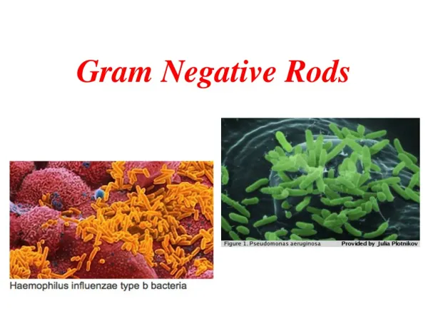
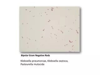
Overview- Haemophilus • Small • Non-motile • Gram-negative rods • Transmitted via respiratory droplets, or direct contact with contaminated secretions • Normal flora of the human respiratory tract and oral cavity.
Haemophilus species of clinical importance 1. H. influenzae -type b is an important human pathogen 2. H. ducreyi -sexually transmitted pathogen (chancroid) 3. OtherHaemophilusare normal flora - H. parainfluenzae – pneumonia & endocarditis - H. aphrophilus – pneumonia & endocarditis - H. aegyptius – pink eye (purulent conjunctivitis)
Differentiation of Species Growth Factor Hemolysis X Y
Haemophilus influenzae • IsoVitaleX-enriched chocolate agar • Requires 2 erythrocyte factors for growth: X (hemin) and V (NAD). • X & V factors are released following lysis of red blood cells • 5% CO2 enhances growth
Public Health Aspects-H. influenzae • Typing based on capsule polysaccharide a → f • Polyribose-ribitol phosphate (PRP) capsule (type b) • Nonencapsulated (nontypeable) organisms are part of normal flora of the respiratory tract • 95% of invasive disease caused by type b
Public Health Aspects • H. influenzae type b incidence has fallen 99% post-vaccine • Pre-immunization • Serotype b was the most common invasive species
Post-immunization • Most cases in unvaccinated or incompletely vaccinated children. • Non-encapsulated and serotype f are the most common • Children - Pneumonia and meningitis less common • Most infections (~2/3) are currently attributed to nontypeable strains.
Disease caused by H. influenzaeSerotype b Clinical Microbiology Reviews, April 2000, p. 302-317, Vol. 13, No. 2
Invasive Diseases post-immunization • Septic arthritis • Osteomyelitis • Cellulitis • Pericarditis • Pneumonia - most frequent is serotype f • Otitis media • Streptococcus pneumoniae and then non-typeable Hi
Pathogenic Mechanisms • H. influenzae • Antiphagocyticpolysaccharide capsule is the major pathogenesis factor • Lipopolysaccharide lipid A component from the cell wall (major role in non capsule strains) • All virulent strains produce neuraminidase and an IgA protease • No exotoxins
Pathogenesis – Host Factors • Hib conjugate vaccine (PRP capsule) • The Hib conjugate vaccine does not protect against nontypeable strains. • Persons at risk for invasive H influenzae disease • Asplenia • Immunocompromised
Public Health Aspect of other Haemophilus strains • H. ducreyi • Sexually transmitted disease - chancroid • H. influenzae biogroup aegyptius • Brazilian Purpuric Fever • H. aegyptius • “pink eye” (purulent conjunctivitis) • H. aphrophilus • pneumonia • Infective endocarditis
Haemophilus ducreyi- chancroid • ~5,000 cases per year in the US
Haemophilus ducreyi • Occurs in strands • Grows of chocolate agar requires factors X (hemin) but not factor V (NAD)
Haemophilusinfluenzaebiogroupaegyptius • Brazilian purpuric fever in children • High fever • Death within 48 hours
Antimicrobial therapy • Ampicillin has historically been the drug of choice for serious invasive H. influenzae infections, especially meningitis • Since the mid-1980s 10 to 20% of Haemophilus species have become ampicillin resistant (plasmid mediated beta lactamase) • Chloramphenicol was used as the “back-up” antibiotic for ampicillin resistant strains • Chloramphenicol resistant strains have now begun to appear • Third generation cephalosporins are now the drugs of choice • Also Trimethoprim-sulfamethoxazole or ciprofloxicin are clinically effective for strains that are ampicillin and chloramphenicol resistant
Antimicrobial testing • All isolates from blood or CSF, and isolates involved in life threatening infections are tested for beta lactamase • The most widely used test employs a chromogenic cephalospirin substrate, Cefinase • If the isolate does not produce beta lactamase (negative test result) ampicillin should be effective • If the beta lactamase test is positive a modified Kirby Bauer disk diffusion test is performed utilizing a Haemophilus test medium (HTM) that contains X & V rather than the regular Kirby Bauer medium, Mueller-Hinton. • The Kirby Bauer test determines extent of resistance – ie. MIC of antimicrobial agent, in this case ampicillin
Antimicrobial testing • The E-test on HTM agar can also be used to test for antimicrobial susceptibility of Haemophilus • The E-test uses a plastic strip that is impregnated with a concentration gradient of antimicrobial agents • The strip is placed on a plate of HTM agar that has been spread-plate inoculated with a 0.5 MacFarland broth culture • The antimicrobial gradient diffuses into the agar • Susceptible strains forms an elliptical pattern of inhibition • The point near the low concentration end of the strip that is intersected by the zone of inhibition represents the minimal inhibitory concentration (MIC) of the antimicrobial agent
E-Test E High concentration end of the strip Zone of Inhibition O.50ug/ml line Bacterial Growth Low concentration end of the strip
LEGIONELLAEOverview • Facultative intracellular pathogen • Gram negative rod • Requires specialized media to grow • Stains poorly with gram stain • Transmitted via contaminated aerosols • No person to person transmission
2Species of Clinical Importance • Legionella • One genus • 50 species • ½ of species implicated in human disease • Legionellapneumophila • Causes ~ 90% of all cases of legionellosis • Majority of all confirmed cases are caused by serogroups 1-6 • Legionellamicdade • Most common after L. pneumophila
Legionellamicdadei • Caution: • This strain can stain weakly acid fast on primary isolation, but loses this property when grown in vitro. • NO RELATIONSHIP TO MYCOBACTERIA
Microbiology • Will not grow on standard Sheep Blood Agar • Buffered Charcoal Yeast Extract Agar (BCYE) • 1. Cysteine is essential for growth • Iron is essential for growth • Growth conditions: • 350 C • 3-7 days
Colony Appearance: • Ground glass • Small 1-3 mm
Gram staining show Legionella are poorly staining, slender, rods © 2005 Elsevier
Laboratory Diagnosis of Legionella • Culture of Legionella organism from normally sterile tissue • Detection of L. pneumophilaantigen in urine • Seroconversion: 4 fold or greater rise in specific serum antibody titer L. pneumophila • Direct fluorescent antibody (DFA) staining
Legionnaires disease- Public Health • Disease - Worldwide • Sporadic • Epidemic community-acquired pneumonia • Nosocomial infections • Exposure - Water-based aerosols • Air conditioning cooling towers • Whirlpool spas • sauna or mister • Survival – Environment • Amoebae • biofilms
2 Clinical Presentations • Legionnaire's disease • Incubation period 2-10 days • pneumonia • 15-75% mortality • erythromycin • Pontiac fever • Incubation period 1-2 days • flu-like • milder (no mortality) • self-limiting
Pathogenesis of Legionella • Phagocytosis into the monocytes • binding to complement receptors • Inhibition of phagolysosome fusion • Replication within the phagosome • Lysis of the phagosome leads to apoptosis and release of the organism • TH1 cells and IFN-γ
Bordetella pertussis • Strict aerobe • Gram negative • Small Coccobacillus -singly or in pairs • Transmission by aerosolized droplets • Non-invasive • Strictly human pathogen
B. pertussis Small, transparent hemolytic colonies on Bordet-Gengou medium
Diagnosis • Based on symptoms • Culture of respiratory secretions on Bordet-Gengoumedium • Direct fluorescent antibody testing • PCR • Slide agglutination
Pertussis Pathogenesis • Two-stage process of disease • Respiratory colonization • 7-10 days • NO symptoms • Positive cultures toward the end of this stage • Toxin-mediated disease
Colonization • Fimbriae are NOT involved. • Attachment requires 2 factors • Pertussis Toxin • Filamentous hemagglutinin
Pertussis- Disease • Inflammation interferes with clearance of pulmonary secretions • Cough progresses from mild (catarrhal stage) to sever (paroxysmal stage) • Resolves slowly catarrhal stage Mild coughing, sneezing, or runny nose. paroxysmal stage uncontrollable fits, each with five to ten forceful coughs, followed by a high-pitched "whoop" sound in younger children, or a gasping sound in older children, as the patient struggles to breathe in afterwards
Pertussis- Disease • Primarily a toxin-mediated disease
Pertussis toxin (pertussigen) • Pertussis toxin is an oligopeptide AB-type exotoxin that is the major cause of pertussis (abnormal cough). • T cell lymphocytosis and has adjuvant properties. • hypoglycemia, increased IgE synthesis, and increased histamine and endotoxin sensitivity. • inhibits many leukocyte functions, including chemotaxis, phagocytosis and respiratory burst and impairs NK cell killing • contributes to bacterial binding to ciliated epithelial cells. • covalent addition of ADP-ribose to the GTP binding Gi protein and thereby preventing the deactivation of adenylatecyclase. This results in the accumulation of large amounts of cAMP which leads to increased mucus secretion and interferes with many cellular functions.
Pertussis Toxin AB-toxin (6 protein subunits)
Adenylatecyclase toxin This exotoxin penetrates the host cells, is activated by calmodulin and catalyzes the conversion of ATP to cAMP. Like pertussigen, it also inhibits phagocyte and NK cell functions. However, in contrast with pertussigen, the cAMP increase caused by this toxin is short-lived The increase in cAMP leads to: 1. Inhibition of chemotaxis 2. Inhibition of phagocytic activity 3. Inhibition of superoxide generation 4. Induction of apoptosis in macrophages
Tracheal cytotoxin This is a peptidoglycan-like molecule (monomer) which binds to ciliated epithelial cells, thus interfering with ciliary movement. In higher concentrations, it causes ciliated epithelial cell extrusion and destruction. The destruction of these cells contributes to pertussis. Dermonecrotic(heat-labile) toxin Dermonecrotic toxin is a very strong vaso-constrictor and causes ischemia and extravasation of leukocytes and, in association with tracheal cytotoxin, causes necrosis of the tracheal tissue. Filamentous haemagglutinins (agglutinogens) These are not exotoxins but are filament-associated lipo-oligo-saccharides which are implicated in the binding of the organism to ciliated epithelial cells. Antibodies against these molecules are protective, probably by preventing bacterial attachment.