Download

1 / 68
690 likes | 839 Vues
Explore the intricate process of heart development from formation to twisting and partitioning, detailing the crucial stages and structural transformations that shape the heart. Unravel the mysteries behind the formation of valves and division of the heart halves.

E N D
Development of the heart Mark Kozsurek, M.D., Ph.D. mark@kozsurek.hu ED I., 20/02/2018
Development of theheartincludes: • formation of thehearttube, • twisting and furtherdifferentiation of thehearttube, • partitioning of theheart – completeisolationoftheleft and right halves, • formation of thevalves.
I. Early development of the heart: appearance of the heart tube
oropharyngeal membrane cloacal membrane amniotic cavity yolk sac extraembryonic celom septum transversum connecting stalk cardiogenic area
Note that heart starts to develop anterior to the brain! brain spinal cord
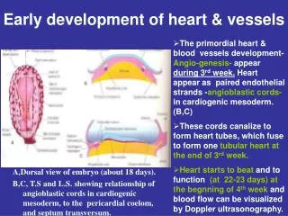
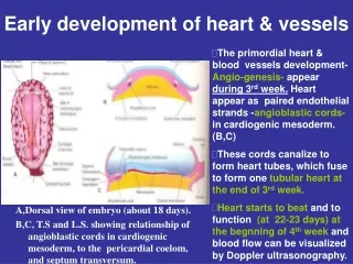
Heart primordium appears on the 18th-19th days (the end of the third week). At this stage the embryo is a flat, trilaminar disc and the differentiation of the three germ layers has just begun. Heart develops from the cardiogenic area, a part of the intraembryonic mesoderm anterior to the oropharyngeal membrane and posterior to the septum transversum.
lateral plate mesoderm somatic layer splanchnic layer intraembryonic celom amniotic cavity yolk sac cardiogenic area
angioblastic cords somatic layer somatic layer splanchnic layer splanchnic layer splanchnic layer A group of cells leaves the splanchnic mesoderm, moves medially and forms the angioblastic cords.
myoepicardial mantle somatic layer splanchnic layer endocardial tubes On the dorsal side of the endocardial tube, the splanchnic mesoderm thickens and forms the myoepicardial mantles.
Further development of the heart is mainly determined by the folding of the embryo (4th week). The folding includes: 1) „flexion” around transverse axes – this will explain, how the heart primordium moves from „above the head” into the chest occupying its final position. 2) „rolling” around the longitudinal axis – this will mainly explain the fusion of the structures originally developing on the two sides of the embryo.
foregut dorsal mesocardium intraembryonic celom pericardial cavity The „rolling”
Folding of the embryo, especially the „rolling”, results in the fusion of the two endocardial tubes in the midline. Somewhat later the myoepicardial mantles also unite and surround the endocardial tube. The intraembryonic celoms fuse ventrally, and finally, the dorsal mesocardium disappears.
The heart tube, branching both rostrally and caudally, is surrounded by the pericardial cavity.
Summary • Cellsemigratingfromthesplanchnicmesodermformtheendocardialtubes, thisgiveslatertheENDOCARDIUM. • Thickening of thesplanchnicmesodermgivesthemyoepicardialmantlesfromwhichthe MYOCARDIUM, and theVISCERAL LAYER OF THE PERICARDIUM (EPICARDIUM) develop. • Intraembryoniccelom, originallyboundedbythelateralplates, transformsintothe PERICARDIAL CAVITY. • Somaticlayer of themesodermdifferenciatesintothe PARIETAL LAYER OF THE PERICARDIUM.
As the two ends of the heart tube are fixed, its intense longitudinal growth necessarily results in the formation of several loops and curvatures. Furthermore some parts of the tube remain narrower, while others dilate. The venous end moves backward and ascends and gets behind the slightly descending arterious end. cardiogenic plate endocardial tubes and their fusion 6 5 4 3 1-2 6 6 • sinus venosus • commonatrium • atrioventricularcanal • commonventricle • bulbuscordis • truncusarteriosus 4 5 4 3 5 RV 3 2 LV 2 1 1 Common ventricle gives the later LV and the inflow part of the RV, while the bulbus cordis differentiates into the outflow part of the RV!
sup. post. ant. inf. truncus arteriosus sinus venosus TRANSVERSE SINUS common atrium bulbus cordis atrioventricular canal common ventricle OBLIQUE SINUS
left sinus horn sinus venosus right sinus horn sinu-atrial opening truncus arteriosus sinusvenosus common atrium common atrium bulbus cordis atrioventricular canal lateral view atrioventricular canal Note the right and left sinus horns draining into the sinus venosus and the right and left veinous valves (green) at the sinu-atrial opening! common ventricle common ventricle frontal view
sinus venosus left sinus horn → coronary sinus truncus arteriosus right sinus horn sinusvenosus sinu-atrial opening bulbus cordis common atrium common atrium common ventricle atrioventricular canal atrioventricular canal The sinu-atrial opening moves to the right, the left sinus horn (or duct of Cuviér) involutes and persists as the coronary sinus. common ventricle
SVC left sinus horn → coronary sinus truncus arteriosus sinus venarum sinusvenosus sinu-atrial opening bulbus cordis common atrium IVC common atrium common ventricle atrioventricular canal atrioventricular canal Note the three veins opening into the sinus venarum (derivative of the sinus venosus): the superior and inferior vena cava and the coronary sinus! common ventricle
SVC pulmonaryveins coronary sinus truncus arteriosus sinus venarum sinusvenosus sinu-atrial opening bulbus cordis common atrium IVC common atrium common ventricle atrioventricular canal atrioventricular canal Pulmonary veins arise from (or enter) the left portion of the common atrium. common ventricle
SVC pulmonaryveins coronary sinus truncus arteriosus sinus venarum sinusvenosus sinu-atrial opening bulbus cordis common atrium IVC common atrium common ventricle atrioventricular canal SMOOTH ROUGH atrioventricular canal Sinus venarum and pulmonary vein segments getting incorporated into the atrial wall will give the smooth-walled parts of the atria (blue), while the original regions of the common atrium will persist as the rough parts of the atria. common ventricle
Septation of the common atrioventricular (AV) orifice. • Formation of the interatrial septum. • Formation of the muscular interventricular septum. • Appearance of the membranous interventricular septum and the spiral aorticopulmonary septum.
septum primum septum primum
septum primum septum primum
septum primum septum primum foramen primum foramen primum
septum primum septum primum foramen primum foramen primum
septum primum septum primum foramen secundum foramen secundum foramen primum foramen primum
septum primum septum primum foramen secundum foramen secundum foramen primum foramen primum
septum primum septum primum foramen secundum foramen secundum
septum secundum septum secundum foramen secundum foramen ovale septum primum foramen ovale septum primum
septum secundum septum secundum foramen secundum foramen ovale septum primum foramen ovale septum primum
septum secundum septum secundum foramen secundum septum primum foramen ovale foramen ovale septum primum
Summary • The septation of the common atrium starts with the appearance of the crescent-shaped septum primum. The opening of this septum, the foramen primum, becomes progressively smaller. • Before the foramen primum completly closes, postero-superiorly several small openings appear on the septum primum. These perforations coalesce later and form the foramen secundum. • On the right side of the septum primum a new septum, the septum secundum, starts to grow. The orifice of the septum secundum is the foramen ovale. • Finally two crescent-like, incomplete, partially overlapping septa exist with one hole on each. Septum secundum is more rigid and the septum primum on its left side acts as a valve letting the blood flow exclusively from the right to the left. Its importance will become obvious when the fetal circulation is discussed.
septum secundum septum secundum foramen secundum septum primum foramen ovale foramen ovale septum primum
septum secundum septum secundum foramen secundum septum primum foramen ovale foramen ovale septum primum
septum secundum interventricular foramen foramen ovale septum primum septum secundum foramen secundum septum primum foramen ovale interventricular foramen
Summary • Nothing is simplier then the development of the muscular part of the interventricular septum. • Similarly to the processes observeble at the level of atria, a crescent-like septum appears on the floor of the common ventricle. This septum grows upward until it reaches the bulbus cordis. Here the interventricular foramen persists for a while and will be closed by an other septum.
dorsal aortae septum secundum truncus arteriosus 6th aortic arches * interventricular foramen foramen ovale septum primum bulbus cordis *: differentiate into pulmonary arteries!!!
6th aortic arches dorsal aortae In the bulbus cordis and the truncus arteriosus two endocardial thickenings, bulbar and truncal ridges appear and exhibit a spiral course twisting 270 degrees* (90 degrees in each third of the full length). * Different values between 180 and 270 degrees can be found in different textbooks.
6th aortic arches dorsal aortae The two bulbar and truncal ridges (= conotruncal ridges as used by others) approach each other.
„hook” 6th aortic arches dorsal aortae Finally, a complete spiral septum is formed. An additional hook-shaped septum arises from the superior edge of this septum and isolates the posterior portion of the truncus arteriosus.
dorsal aortae pulmonary arteries Venous blood (blue) from the right ventricle ascends and from the posterior portion of the truncus arteriosus may only flow toward the pulmonary arteries developing from the 6th aortic arches. Blood from the left ventricle (red) exits through the two dorsal aortae.
dorsal aortae pulmonary arteries septum secundum foramen ovale interventricular foramen septum primum
dorsal aortae septum secundum pulmonary arteries foramen ovale septum primum