Heart Development Lecture by Prof. Saeed Abuel Makarem
Learn about the intricate process of heart tube formation and development, including common anomalies. Explore the fate of the heart tube and its transformation into a vital organ.

Heart Development Lecture by Prof. Saeed Abuel Makarem
E N D
Presentation Transcript
HEART DEVELOPMENT By Prof. Saeed Abuel Makarem
Objectives • By the end of the lecture you should be able to: • Describe the formation, site, union, division of the of the heart tube. • Describe the formation and fate of the sinus venosus. • Describe the formation of the interatrial and the interventricular septae. • Describe the formation of the two atriaand the two ventricles. • Describe the partitioning of the truncus arteriosus and formation of the aorta and pulmonary trunk. • List the most common cardiac anomalies.
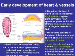
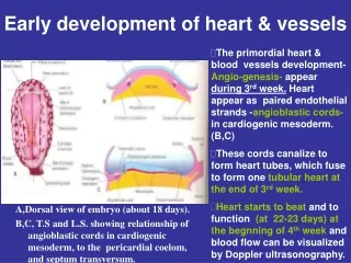
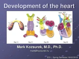
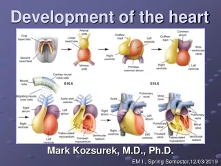
FORMATION OF THE HEART TUBE • The heart is the first functional organ to develop. • It develops from splanchnic mesoderm (cardiogenic area), cranial to the developing mouth & nervous system and ventral to the developing pericardial sac. • The heart primordium is first evident at 18 days(as an angioplastic cords which soon canalize to form the 2 heart tubes). • As the head fold completed, the developing heart tubes lie in the ventral aspect of the embryo dorsal to the developing pericardial sac. • After lateral folding of the embryo • The 2 heart tubes fuse together to form a single endocardial heart tube. • It begins to beat at 22 to 23 days.
Blood flow begins during the beginning of the fourth week and can be visualized by Ultrasound Doppler
Development of the Heart tube • After lateral folding of the embryo, the 2 heart tubes approach each other and fuse to form a single endocardial heart tube within the pericardial sac. • Fusion of the two tubes occurs in a craniocaudaldirection.
What is the fate of the Heart Tube? • The heart tube grows faster than the pericardial sac, so it shows 5 alternate dilations separated by 4 constrictions. • These are: • Sinus Venosus. • Truncus Arteriosus. • Bulbus Cordis. • Common Ventricle. • Common Atrium. The endocardial heart tube has 2 ends: 1. Venous end; Sinus Venosus. 2. Arterial end; Truncus arteriosus
U-SHAPED HEART TUBE • Bulbus cordis and ventricle grow faster than other regions. • So the heart bends upon itself, forming • The U-shaped heart tube, (bulboventricular loop).
Loop formation Or S-Shaped Heart Tube • As the heart tube more develops it bends, upon itself: SO, the atrium and sinus venosus become dorsal to the truncus arteriosus, bulbus cordis, and ventricle. • By this stage the sinus venosus has developed 2 lateral expansions, called the 2 horns ( right and left horns), and body.
Veins Associated With Heart Development Each horn of the sinus venosusreceives 3 veins: 1.Common cardinal 2.Vitelline 3.Umbilical Cardinal vein from the fetal body. Vitelline from the yolk sac. Umbilical from the placenta.
Fate of Sinus Venosus • The right horn forms the smooth posterior wall of the right atrium. • The left horn and body atrophy and form the coronary sinus. • The left common cardinal vein forms the oblique vein of the left atrium.
Right Atrium • The right horn of the sinus venosus forms the smooth posterior part of the right atrium. • The roughTrabeculated anterior part of the right atrium is derived from the primordial common atrium. • These two parts are demarcated by the crista terminalis internally and sulcus terminalis externally.
Left Atrium • Rough Trabeculated part: derived from the common primordial atrium. • The smooth part: derived from the absorbed Pulmonary Veins.
Partitioning of Primordial Heart Partitioning of: 1- Atrioventricular canal. 2- Common atrium. 3- Common ventricle. 4- Bulbus cordis. It begins by the middle of 4th week. It is completed by the end of 5th week.
Partitioning of the atrioventricular canal • Two endocardial cushions are formed on the dorsal and ventral walls of the AV canal. • The AV endocardial cushions approach each other and fuse to form the septum intermedium. • Dividing the AV canal into right & left canals. • These canals partially separate the primordial atrium from the primordial ventricle.
Partition of the common atrium Septum Primum • It is sickle- shaped septum that grows from the roof of the common atrium towards the fusing endocardial cushions (septum intermedium) • So it divides the common atrium into right & left halves.
Ostium Primum • The two ends of the septum primum reach to the growing endocardial cushions before its central part. • So the septum primum bounds a foramen called ostium primum. • It serves as a shunt, enabling the oxygenated blood to pass from right to left atrium. • The ostium primum become smaller and disappears as the septum primum fuses completely with endocardial cushions (septum intermedium) to form the interatrial septum.
Septum Secundum • The upper part of septum primum that is attached to the roof of the common atrium shows gradual resorption forming an opening called ostium secondum. • Another septum descends on the right side of the septum primum called septum secundum. • It forms an incomplete partition between the two atria. • Consequently a valvular oval foramen forms, (foramen ovale)
Fate of foramen Ovale • At birth when the lung circulation begins the pressure in the left atrium increases and exceeds that of the right atrium. • So the two septae oppose each other. • Its site is represented by the Fossa Ovalis. • The septum primum forms the floor of the fossa ovalis. • The septum secondum forms the margin of the fossa ovalis which also called the limbus (anulus) ovalis.
Partitioning of Primordial Ventricle Muscular part of the interventricular septum. • Division of the primordial ventricle is first indicated by a median muscular ridge, (primordial interventricular septum). • It is a thick crescentic fold which has a concave upper free edge. • This septum bounds a temporary connection between the two ventricles called interventricular foramen.
Interventricular Septum The membranous part of the IV septum is derived from: 1- A tissue extension from the right side of the endocardial cushion. 2- Aorticopulmonary septum. 3- Thick muscular part of the IV septum.
Spiral Aorticopulmonary Septum • A spiral septum develops in the truncus arteriosus dividing it into aorta and pulmonary trunk. • So, now the pulmonary artery joins the rightventricle while the aorta joins the left ventricle.
BULBUS CORDIS • The bulbus cordis forms the smooth upper part of the two ventricles. • In the right Ventricle: • It forms the ConusArteriosus or (Infundibulum) which leads to the pulmonary trunk. • In the left ventricle: • It forms the aortic Vestibule which leads to the aorta.
Atrial Septal Defects (ASD) • Absence of septum primum and septum secundum, leads to common atrium. • Absence of Septum Secundum
Excessive resorption of septum primum(ASD) Patent foramen ovale
VENTRICULAR SEPTAL DEFECT (VSD) • Roger’s disease • Absence of the membranouspart of interventricular septum. • Usually accompanied by other cardiac defects.
TETRALOGY OF FALLOT Blue Baby • Fallot’s Tetralogy: • 1-VSD. • 2- Pulmonary stenosis. • 3-Overriding of the aorta • 4- Right ventricular hypertrophy.
TETRALOGY OF FALLOT Blue Baby
(TGA) OR TRANSPOSITION OF GREAT ARTERIES • TGA is due to abnormal rotation or malformation of the aorticopulmonary septum, so the right ventricle joins the aorta, while the left ventricle joins the pulmonary artery. • It is one of the most common cause of cyanotic heart disease in the newborn. • Often associated with ASD or VSD. Blue Baby
Persistent Truncus Arteriosus • It is due to failure of the development of the aorticopulmonary (spiral) septum. • It is usually accompanied with VSD.