Download
1 / 1
20 likes | 271 Vues
P13.24 Longitudinal study of uterine artery Doppler after normal vaginal delivery. A. Mulic-Lutvica, K. Eurenius, O. Axelsson Institution of Women’s and Children’s Health, Uppsala University Hospital. Introduction

E N D
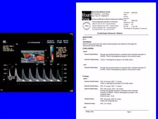
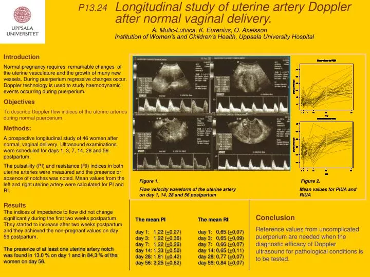
P13.24 Longitudinal study of uterine artery Doppler after normal vaginal delivery. A. Mulic-Lutvica, K. Eurenius, O. Axelsson Institution of Women’s and Children’s Health, Uppsala University Hospital Introduction Normal pregnancy requires remarkable changes of the uterine vasculature and the growth of many new vessels. During puerperium regressive changes occur. Doppler technology is used to study haemodynamic events occurring during puerperium. Objectives To describe Doppler flow indices of the uterine arteries during normal puerperium. Methods: A prospective longitudinal study of 46 women after normal, vaginal delivery. Ultrasound examinations were scheduled for days 1, 3, 7, 14, 28 and 56 postpartum. The pulsatility (PI) and resistance (RI) indices in both uterine arteries were measured and the presence or absence of notches was noted. Mean values from the left and right uterine artery were calculated for PI and RI. Figure 1. Flow velocity waveform of the uterine artery on day 1, 14, 28 and 56 postpartum Figure 2. Mean values for PIUA and RIUA Results The indices of impedance to flow did not change significantly during the first two weeks postpartum. They started to increase after two weeks postpartum and they achieved the non-pregnant values on day 56 postpartum. The presence of at least one uterine artery notch was found in 13.0 % on day 1 and in 84,3 % of the women on day 56. The mean PIThe mean RI day 1: 1,22 (+0,27) day 1: 0,65 (+0,07) day 3: 1,22 (+0,36) day 3: 0,65 (+0,09) day 7: 1,22 (+0,26) day 7: 0,66 (+0,07) day 14: 1,33 (+0,50) day 14: 0,65 (+0,11) day 28: 1,81 (+0,42) day 28: 0,77 (+0,07) day 56: 2,25 (+0,62) day 56: 0,84 (+0,07) Conclusion Reference values from uncomplicated puerperium are needed when the diagnostic efficacy of Doppler ultrasound for pathological conditions is to be tested.