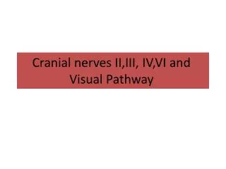
Understanding the Primary Visual Pathway in Cognitive Neuroscience
Explore the primary visual pathway from the eyes to the brain's visual cortex, delving into information processing stages and experimental strategies to uncover mechanisms of visual perception. Learn about key figures like David Hubel and Thorsten Wiesel and their contributions to our understanding of visual information processing. Dive into topics like photoreceptor function, receptive fields of visual neurons, and color sensitivity in retinal ganglion and LGN neurons. Discover the functional significance of center-surround fields and how they enhance contrast perception and boundary detection. Join us in unraveling the complexities of visual processing in the brain.

Understanding the Primary Visual Pathway in Cognitive Neuroscience
E N D
Presentation Transcript
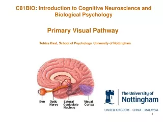
C81BIO: Introduction to Cognitive Neuroscience and Biological Psychology Primary Visual Pathway Tobias Bast, School of Psychology, University of Nottingham
Today’s lecture Next lecture Initial stages of visual information processing – from retina to V1 (primary visual pathway) Visual perception, memory, etc. – beyond V1 (focus on occipito-temporal pathway)
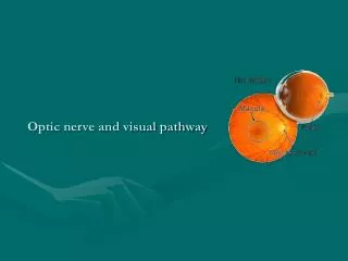
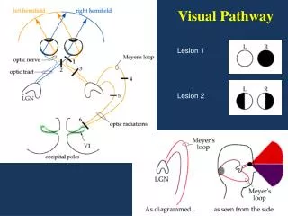
Primary visual pathway from eyes to primary visual cortex (striate cortex, V1) in the occipital lobe Right half of field of view Left half of field of view Central 1o Optic nerve Optic chiasm Optic tract Retina Lateral Geniculate Nucleus (LGN) Fovea
Information-processing stages in primary visual pathway Visual cortex Lateral geniculate body Retina Optic nerve Retinal ganglion cells Bipolar cells Photo-receptors Experimental strategy to reveal mechanisms of visual perception By studying the different neuronal responses at different stages of the visual pathway, one may gain understanding of the different stages of visual information processing that mediate visual perception.
Experimental set-up to record visual responses of neurons along the visual pathway
Seminal contributions to our understanding of visual information processing David Hubel Thorsten Wiesel Nobel Prize in Physiology or Medicine 1981 For discoveries concerning information processing in the visual system http://nobelprize.org/nobel_prizes/medicine/laureates/1981/index.html The neurobiology group in the Department of Pharmacology at Harvard Medical School, 1963, the group that later formed the Department of Neurobiology.
Information-processing stages in primary visual pathway Visual cortex Lateral geniculate body Retina Optic nerve Retinal ganglion cells Bipolar cells Photo-receptors
Photoreceptors • Rods -More abundant (ca. 120 million in human retina) -No colour (i.e., wave length) discrimination -Sensitive in low light levels -Higher density in periphery (don’t look directly at dim stars) -Track high-rate changes (see flicker of 60Hz monitor from corner of your eyes) Back of eye • Cones -Less abundant (ca. 6 million in human retina) -3 types discriminate different wavelengths (S,M,L) -Less sensitive to low light -Higher concentration in fovea -Cannot follow rapid changes (can’t see 60-Hz flicker when directly looking at monitor) Photoreceptors and bipolar cells vary their voltage as theyare stimulated (analogue signal), whereas all subsequent cells vary spike rate (all-or-nothing, digital signal). Rod Cone Photoreceptor detection of light is translated into excitation or inhibition of retinal ganglion cells via bipolar cells.
Life without cones or rods: • Without cones we could only see in shades of black and white. • Without rods we could only see in shades of black and white. • Without cones we would have more trouble seeing things in the dawn. • a) and c) are correct.
Receptive fields of visual neurons • The portion of the retina/visual field in which visual stimulation will evoke a change in the firing rate of a given visual neuron. • Substructure of a receptive field: A description of how visual stimuli need to be presented in the receptive field of a visual neuron in order to evoke firing-rate changes.
Retinal ganglion neurons - - + - - + + - + + • Receive input from multiple photoreceptors (via bipolar cells) • ON-OFF Centre-Surround receptive fields Present stimulus ON centre – OFF surround • Light presented in ‘ON’ regions excites cell, and light in ‘OFF’ regions inhibits cell • ON and OFF regions are organised in ‘centre-surround’ fashion • Response rate of cell is based on the sum of stimulation in ON region minus stimulation in OFF region OFF centre – ON surround Enhancement of contrast and boundaries Neurons in the lateral geniculate body respond to visual stimuli in similar ways to retinal ganglion cells.
Functional significance of centre-surround fields • The world has lots of things that stay constant, and we don’t need to keep responding to them – what counts most are changes and boundaries. So, responding only to changes and boundaries (edges) is efficient. • The luminance of features is represented relative to their surround. This helps preserve appearance of objects regardless of light levels in the environment (newspaper looks basically the same in a dark room and in sunlight, despite hugely different levels of overall reflected light). However, it can also result in illusions:
See a video demonstrating the eye camera at: ScientificAmerican.com/oct2012/dvs Paper by Sejnowski & Delbruck (2012, Scientific American) available from Moodle or my webpage
Colour sensitivity of retinal ganglion and LGN neurons • Retinal ganglion and LGN cells receive inputs from cones (that are differentially sensitive to different wavelengths) and are sensitive to colour • Colour-sensitive retinal ganglion and LGN neurons have receptive fields that show centre-surround colour opponency + + + + - - - - Functional significance of colour-opponency not clear. However, colour opponency, together with firing-rate adaptation (rebound effects), in retinal ganglion cells can explain negative afterimages.
Life without centre-surround receptive fields: • Without an on-off centre-surround organisation of the receptive fields in some neurons of primary visual pathway we’d struggle more to detect contrasts and edges. • We could not distinguish black and white. • We would have more difficulties to recognize objects if light levels in the environment change. • a) and c) are correct.
Information-processing stages in primary visual pathway Visual cortex Lateral geniculate body Retina Optic nerve Retinal ganglion cells Bipolar cells Photo-receptors
Primary visual cortex (striate cortex, V1) Human Calcarine fissure Macaque V1
Orientation-selective cells in V1 Simple cells • Fields have inhibitory and excitatory regions. -Can be thought of as combining inputs from ON and OFF cells. Complex cells • Most V1 neurons respond to elongated stimuli with specific orientation. • Two main types of orientation-sensitive V1 neurons • Fields have no discrete ON and OFF regions. • Best response to moving stimuli (reflecting response adaptation). -Can be thought of as combining inputs from simple cells.
Stimulate your simple and complex cells in V1 Download CEMIT Lite from the App Store.
Maps and modules in V1 Modules Retino-topic map V1 is divided into small columnar modules that combine neurons sensitive to different aspects of stimuli presented in a small part of the visual field. Orderly mapping of retina/visual field onto visual cortex Right retina/visual field
Further processing of visual information • To result in perception and memory of the ‘holistic’ visual properties of whole objects and visuals scenes, the visual information from the modules in V1 needs to be combined and further processed. • This processing takes place in the visual association cortices (V2-V5, inferior temporal cortex, posterior parietal cortex) and other regions. NEXT LECTURE!!
Blindsight • Subjects with lesions to primary visual cortex and apparent ‘blindness’ can show appropriate responses to visual stimuli of which they are not ‘conscious’. • Examples of such ‘blindsight’ include: ‘looking’ (i.e., moving the eyes) or pointing toward visual stimuli; detection of movement; etc. • ‘Blindsight’ highlights that, apart from the primary visual pathway that is critical for conscious vision, there are additional visual pathways. • Recent study suggests that direct LGN projections to extrastriate cortex are critical for blindsight (Schmid et al., 2010, Nature 466:373-377). • ‘Blindsight’ also highlights that the brain can perform visual information processing which can guide subjects’ behaviour without their conscious awareness. Cowey & Stoerig (1991) The neurobiology of blindsight. Trends Neurosci. 14:140-145.
Primary visual pathway – Selected Reading Textbook chapter: Carlson NR (any recent edition) The physiology of behavior. Chapter 6, Vision. Excellent book (many figures used in lecture come from this book): Hubel D (1995) Eye, brain, and vision. Scientific American Library/Scientific American Books. (Available in George Green Library). Review article: Hubel D, Wiesel T (1998) Early explorations of the visual cortex. Neuron 20:401-420.
Primary visual pathway – Some questions to guide revision • Which brain regions make up the primary visual pathway? • What is a visual receptive field, what is its substructure? • Can you outline key stages of visual information processing along the primary visual pathway, characterizing the properties of neurons at different stages by describing the neurons’ visual receptive fields? • Can you give a physiological explanation of negative afterimages? • What is ‘Blindsight’ and what are the conceptual implications of this phenomenon?
Primary visual pathway – Some questions to think about • How could you explain these selective visual field defects (that can occur in neurological patients) based on a diagram of the visual pathway? • Which processing stages could lead to the perception of or ? • Do we “see” the world as it is? (eyes are tested one after the other, with one eye shut)