Eye &Visual Pathway
Eye &Visual Pathway. Dr. Nimir Dr. Safaa. Ojectives Describe the location of the orbit. Make a list of structures making the orbit starting from orbital margin. Define each component. Describe openings into orbital cavity.


Eye &Visual Pathway
E N D
Presentation Transcript
Eye &Visual Pathway Dr. Nimir Dr. Safaa
Ojectives • Describe the location of the orbit. • Make a list of structures making the orbit starting from orbital margin. • Define each component. • Describe openings into orbital cavity. • Describe muscles of the orbit, their cone arrangement, origin, insertion, nerve supply and their function. • Describe the visual pathway • Discuss the central connections of the cranial nerves (III,IV,VI)
The Orbital Region • The orbits are a pair of bony cavities • It contains: • The eyeballs &their associated muscles • Nerves, vessels, and fat • Most of the lacrimal apparatus. • .
The Orbit • Description • It is a pyramidal cavity with its base in front and its apex behind. • The orbital margin is formed: Superiorly: Frontal bone • Laterally: Processes of the frontal and zygomatic bones • Inferiorly: zygomatic bone and the maxilla, • Medially :the processes of the maxilla and the frontal bone.
The orbital walls : • Roof: Formed by the orbital plate of the frontal bone • Lateral wall: Formed by the zygomatic bone and the greater wing of the sphenoid • Floor: Formed by the orbital plate of the maxilla • Medial wall: Formed from before backward by the frontal process of the maxilla, the lacrimal bone, the orbital plate of the ethmoid
Eyelids: • The orbital opening is guarded by eyelids • Upper & lower eyelid meet each other at medial and lateral angles. • The palpebral fissure is elliptical opening between eyelids. • Superficial surface of eyelids is covered by skin, and deep surface is covered by a mucous membrane, called the conjunctiva.
Eyelids are formed by orbital septum (fibrous sheet),which is attached to periosteum at orbital margins. • Orbital septum is thickened at margins of lids to form superior and inferior tarsal plates. • Lateral & medial ends of the plates are attached by lateral palpebral ligament & medial palpebral ligaments to orbital bones. • Tarsal glands are long, modified sebaceous glands that secrete oily sustance onto margin of eyelid. • They are embedded in tarsal plates .
Eyelashes are short, curved hairs on free edges of eyelids at mucocutaneous junction. • The conjunctiva is a thin mucous membrane that lines eyelids and is reflected onto anterior surface of eyeball. Its epithelium is continuous with that of cornea. • Sebaceous glands (glands of Zeis) open directly into eyelash follicles. • The ciliary glands (glands of Moll) are modified sweat glands that open separately between adjacent lashes.
Lacrimal Gland: • It consists of a large orbital part and a small palpebral part & situated in superolateral region of the orbit. • It has about 12 ducts which empty tears into lateral part of superior fornix of the conjunctiva.
Tears enter lacrimalcanaliculiwhich open into lacrimal sac then to nasolacrimal duct. • Nasolacrimal duct is about 0.5 in. (1.3 cm) long. • It descends in a bony canal and opens into inferior meatus of the nose.
Openings into orbital cavity: • Superior orbital fissure: • It communicates with middle cranial fossa. • It transmits,lacrimalnerve, frontal nerve, trochlear nerve, abducent nerve, nasociliary nerve, and superior ophthalmic vein. • Inferior orbital fissure communicates with pterygopalatinefossa. • It transmits maxillary nerve and its zygomatic branch, inferior ophthalmic vein, and sympathetic nerves.
Optic canal: • It communicates with middle cranial fossa. • It transmits optic nerve and ophthalmic artery. • Supraorbital notch (Foramen) for supraorbital nerve and blood vessels. • Infraorbital groove and canal for infraorbital nerve and blood vessels. • Nasolacrimal canal communicates with inferior meatus of nose & transmits the nasolacrimal duct.
Blood Vessels • Ophthalmic Artery: • The ophthalmic artery is a branch of the internal carotid artery. • Branches: • The central artery of the retina. • The muscular branches • The ciliary arteries • The lacrimal artery • The supratrochlear and supraorbital arteries
Ophthalmic Veins: • The superior ophthalmic vein communicates with the facial vein . • The inferior ophthalmic vein communicates through the inferior orbital fissure with the pterygoid venous plexus. • Both veins through the superior orbital fissure and drain into the cavernous sinus.
There are six voluntary muscles: • Levatorpalpebraesuperioris • Superior rectus • Inferior rectus • Medial rectus • Lateral rectus • Superior oblique • Inferior oblique.
Contents of the Eyeball: • Aqueous humor which secreted from ciliary processes, enters posterior chamber then flows into anterior chamber through the pupil and is drained away into canal of Schlemm. • Vitreous body fills the eyeball behind the lens and it supports lens and retina. • Lens is a transparent, biconvex structure which is situated behind iris and in front of vitreous body.
The eyeball consists of three coats: • 1-Fibrous coat consists of: • Sclera(‘White of the eye'posterior opaque part). • Cornea(anterior transparent part). • 2- Vascular coat consists of: • Choroid. • Ciliary body. • Iris.
3-Nervous coat(Retina).It has macula lutea(area of most distinct vision) & optic disc without rods and cones (blind spot). • It consists of: • Outer pigmented layer. • Inner nervous layer(retina proper) which consists of many layers but most important are: • Rods & cones • Bipolar cells • Axons of ganglion cells
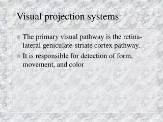
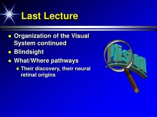
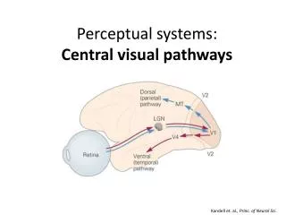
Optic Nerve (Cranial Nerve II): • Fibers of optic nerve are axons of neurons in ganglionic layer of the retina. • They form optic nerve which leaves orbital cavity through optic canal and unites with optic nerve of opposite side to form optic chiasma. • In optic chiasma, nasal (medial) fibers of retina, including the nasal half of macula, cross midline and enter optic tract of opposite side, while temporal (lateral) fibers, including temporal half of macula, pass in same side.
Optic tract passes around cerebral peduncle. Most of its fibers synapse in lateral geniculate body of thalamus. • Few fibers pass to pretectal nucleus and superior colliculusof midbrain for light reflexes. • Axons of geniculate body neurons form optic radiation which terminates in the visual cortex (area 17).