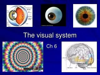
The Visual System: Retinal Mechanisms
160 likes | 285 Vues
This lesson explores the retinal mechanisms involved in processing light stimuli. It covers electromagnetic energy, visible light wavelengths, and the role of photons. We detail the structures involved, including photoreceptors (rods and cones), bipolar cells, and retinal ganglion cells, explaining the transduction process from light reception to action potentials. Key concepts such as receptive fields, center-surround organization, lateral inhibition, and the visual pathway to the primary visual cortex (V1) are highlighted, emphasizing the integration of sensory information for visual perception.

The Visual System: Retinal Mechanisms
E N D
Presentation Transcript
The Visual System:Retinal Mechanisms Lesson 16
The light stimulus • Electromagnetic energy • visible light • 350 – 700 nm (violet – red) • Photon • Wave • Color (hue) - wavelength • Brightness (intensity) – amplitude ~
Color Wavelength Amplitude Brightness
Visual Pathway Optic Nerve Optic Chiasm Optic Tract LGN LGN Lateral Geniculate Nucleus (Thalamus) Optic Radiations V1 V1 Primary Visual Cortex LVF RVF Retina
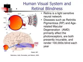
Retinal Organization • Photoreceptors (PR) • transduction • Bipolar cells (BP) • no APs • Retinal ganglion cells (RGC) • 1st APs in pathway • Amacrine & Horizontal cells • communication b/n retinal cells • parallel to retina ~
Retinal Organization Light Which direction does the light enter? PR BP RGC Optic Nerve
Photoreceptors (PR) • Transduction • Rods • achromatic • Cones • color ~
Photoreceptors - PR • Dark current • Depolarized in dark • Na+ influx • NT is released • BP • inhibited ~
Photoreceptors - PR • Light strikes PR • Receptor potential • hyperpolarization • NT release • BP depolarizes • NT release • Excites RGC • APs ~
Rods • Around edges of retina • 120 million • Pigment = rhodopsin • Convergence • high sensitivity • low acuity ~
Cones • Color vision - 3 types • red = long wavelength • green = medium • blue = short • 6 million • Concentrated in fovea • Little convergence • low sensitivity • high acuity ~
Receptive Field (RF) • Region of the retina where... • changes in illumination... • will change the activity of a particular neuron • Center-Surround Organization • BP • RGC • LGN neurons • convergence ~
Center-Surround Organization BP RGC PR
Center-Surround Organization 2 arrangements ON OFF OFF ON
Center-Surround Organization • Light strikes On area of RF • RGC excited APs • Light strikes Off • RGC inhibited APs • Contrast maximal effects on RGC • center & surround lighted cancels out ~
Dark Light PR PR - - H - BP BP + + G G Lateral Inhibition • Center-surround RF • Horizontal cells • no APs • PR stimulates HC • inhibit adjacent BPs • Enhances contrast ~ + Surround Center