Optic nerve and visual pathway
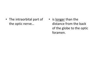
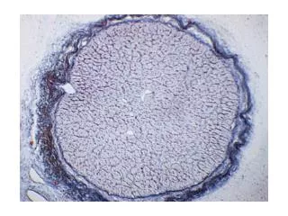
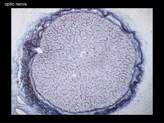
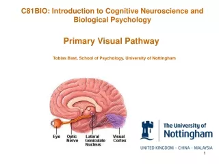
Optic nerve and visual pathway. Optic nerve. The optic nerve is the second cranial nerve situated in the posterior part of the globe It transmits visual impulses from the retina to the brain The head of the optic nerve is called optic disc.
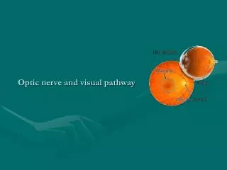

Optic nerve and visual pathway
E N D
Presentation Transcript
Optic nerve • The optic nerve is the second cranial nerve situated in the posterior part of the globe • It transmits visual impulses from the retina to the brain • The head of the optic nerve is called optic disc
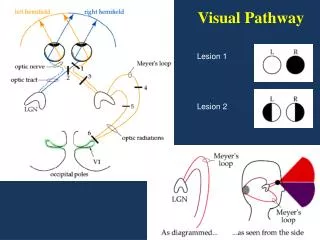
Retinal fibers leave half of the image in opposite side Mutual crossing is known as optic chiasm Nasal fibers mix with uncrossed temporal sector to form optic tract Cell station in brain lateral geniculate body (relay station) Fibers spread out to reach their final destination the visual cortex Visual pathway
Symptoms of optic nerve disease • Loss of vision • Scotomata • Disturbances in color vision • Pain is conspicuously absent, except (retrobulbar)
Signs of optic nerve disease • Relative afferent pupillary defect (RAPD) • Margins, surface, color of the disc • Pulsation of the central retinal vein • Shape and size of the physiological disc
Diseases of optic nerves • Optic neuritis • Neuroretintis • Retrobulbar neuritis • Anterior Ischaemic optic neuropathies (AION) • Papillitis • Optic atrophy
Abnormalities • Drusen : Two kinds of drusen occur in the optic nerve • Common drusen - Disc margins blurred appearance, visual field defect • Giant drusen - Astrocytic hamartomas, that occur with tuberous sclerosis • Conus: It is congenital cresent, choroid and retinal pigment epithelium do not extend upto the optic disc. Defective vision, hypermetropic astigmatism and visual fields defect • Coloboma : Incomplete closure of the embroynic fissure causes optic nerve defects ranging from a deep physiological cup to a pit in the optic disc
AION • It is a segmental infarction of the anterior part of the optic nerve, caused by the occlusion of short posterior ciliary arteries. • Symptoms : Headache, scalp tenderness, jaw claudication, pain and stiffness of the proximal muscles. • Investigation : Fields, ESR, C-reactive protein • Treatment : High dose of oral steroids (tapering dose)
Papillodema • Papillodema is the bilateral non inflammatory passive swelling of the optic disc, produced by raised intracranial tension. • Symptoms: Headache, vomiting without nausea, focal neurologicaldeficit
Optic atrophy • Optic atrophy is the degeneration of optic nerve fibers with loss of their myelin sheaths and characterised by the pallor of the optic disc. • Symptoms : • sudden or gradual loss of field vision • color vision impaired • RAPD • Investigations : • Central field • FFA • X-ray skull • CT scan brain