Connective Tissue Functions and Components
Learn about the diverse functions and components of connective tissue, including the roles of cells, fibers, and ground substances in physical protection, storage, metabolism exchange, repair, and more.

Connective Tissue Functions and Components
E N D
Presentation Transcript
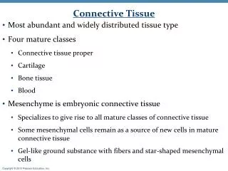
Functions of C.T • Physical and immunologic protection • Storage • Exchange of environmental metabolites • Tissue Repair • Mechanical properties • Binding of different tissues • Lobulation • Defense
Connective tissue: Supporting tissue • A. Types: Diversity based on components • B. Three fundamental components • C. Extra cellular matrix: main part of tissue • D. Embryonic origin: Mesodermal (mesenchyme)
Mesenchymal tissue • Mesenchymal cells - Elongated cells with thin cytoplasmic processes - Oval Nu. - Prominent Nucleoli - Fine chromatin -Multipotential & Undifferentiated cells -Pericytes Or Perivascular cells (Adult stem cells) • An abundant and viscous extracellular substance • Few fibers & lack of collagen
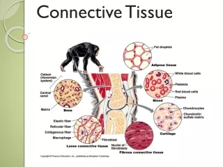
Three fundamental componentsof connective tissue • Three fundamental components: • Cells:Fibroblast/ Macrophage/ Mast cell/Plasma cell/… • Fibers: • Collagen 2. Reticular 3. Elastic • Ground substance: • Glycosaminoglycans (GAGs) & Proteoglycans (PG) • Glycoproteins (GP): fibronectin, laminin,… • Tissue fluid
Simplified representation of the connective tissue cell lineage derived from the multipotential embryonic undifferentiatedmesenchyme cell. Dotted arrows indicate that intermediate cell types exist between the examples illustrated. Note that the cells are not drawn in proportion to actual sizes, eg, adipocyte, megakaryocyte, and osteoclast cells are significantly larger than the other cells illustrated.
Simplified representation of the connective tissue cell lineage derived from the multipotential embryonicundifferentiatedmesenchyme cell. Dotted arrows indicate that intermediate cell types exist between the examples illustrated. Note that the cells are not drawn in proportion to actual sizes, eg, adipocyte, megakaryocyte, and osteoclast cells are significantly larger than the other cells illustrated.
Fibroblast /Fibrocyte -Produce fibers and ECM -Dedifferentiation- wound healing- wound contraction -Myofibroblast during : wound healing/ wound contraction -Growth factors production -Mitosis under specific conditions • Fig. Active (left) and quiescent (right) fibroblasts. External morphologic characteristics and ultrastructure of each cell are shown. Fibroblasts that are actively engaged in synthesis are richer in mitochondria, lipid droplets, Golgi complex, and rough endoplasmic reticulum than are quiescent fibroblasts (fibrocytes)
Medical Application -Regenerative capacity (Reparative capacity) of connective tissue • The main cell type involved in repair is the fibroblast - Dedifferentiation during wound healing: fibrocytes reverts to the fibroblast state - Myofibroblast during wound healing: A cell with features of both fibroblasts and smooth muscle - wound contraction -Mitosis under specific conditions
Fig.6 Section of rat skin. A connective tissue layer (dermis) shows several fibroblasts (F), which are the elongated cells. H&E stain. Medium magnification.
Figure 5—2. Section of rat skin. A connective tissue layer (dermis) shows several fibroblasts (F), which are the elongated cells. Hematoxylin and eosin (H&E) stain. Medium magnification.
Figure 5—3. Quiescent fibroblasts (Inactive fibroblasts) are elongated cells with thin cytoplasmic extensions and condensed chromatin. Pararosaniline-toluidine blue (PT) stain. Medium magnification.
Figure 5—5. Electron micrograph revealing portions of several flattened fibroblasts in dense connective tissue. Abundant mitochondria, rough endoplasmic reticulum (RER), and vesicles distinguish these cells from the less active fibrocytes. Multiple strata of collagen fibrils (C) lie among the fibroblasts. x30,000.
Macrophage and Mononuclear phagocyte system - Origin: Bone marrow --- monocytes-----in connective tissue…..macrophage… ( Monocyte-Macrophage transformation) - Increase in: -Cell size, protein synthesis, number of Golgi and lysosomes - Life span: Up to several months (Long-living cells) - Morphological variation: A wide spectrum of morphological features that correspond to their state of functional activity and to the tissue inhabit
Macrophage and Mononuclear phagocyte system -EM feature: -Developed Golgi, Numerous lysosomes,Microtubules, Microfilaments and RER -10-30 µm - Eccentric, an oval or kidney-shaped Nu. -An irregular surface, protrusions and indentations: (a morphological expression of their active pinocytotic and phagocytic activities )
Macrophage features 1-Fixed macrophage (Histiocyte): - Small & heterochromatin Nu. (fibroblast-like) 2-Free macrophage (activated): - kidney-shaped Nu. + large cytoplasm with residual or phagocytosed bodies - An increase in their capacity for phagocytosis and intracellular digestion - Enhanced metabolic and lysosomal enzyme activity - Removing components during physiologic involution process: uterus involution after parturition
Macrophage functions - Defense functions: - Phagocytose cell debris, neoplastic cells, bacteria, and…. - As an Antigen-presenting cells (APC): Langerhans cells: Dermis - Participate in: 1. Early stages of tissue repair 1.Cell –mediated resistance to infection 2.Cell-mediated resistance to tumors 3. Destruction of aged eryhtrocytes 4. Removing components during physiologic involution process: uterus involution after parturition 5. Tumor cell- killing (capacity)
Macrophage and Mononuclear phagocyte system -Mononuclear phagocyte system - Kuppfer : Liver - Lung macrophage (Dust cell) : Lung - Microglia : CNS - Langerhans : Skin (Dermis) - Osteoclast : Bone -Dendritic cell : Lymph node -Macrophages secretions (secretory cells): 1- Different enzymes : such as Collagenase 2- Cytokines : such as IL-1 & TNF-α
Figure 5—7. Electron micrograph of a macrophage. Note the secondary lysosomes (L), the nucleus (N), and the nucleolus (Nu). The arrows indicate phagocytic vacuoles.
Figure 5—8. Electron micrograph of several macrophages and 2 eosinophils in a region adjacent to a tumor. This figure illustrates the participation of macrophages in tissue reaction to tumor invasion.
Mast cell -General feature: 1-Oval to Round shape 2- 7-20 µm diameter 3- Central Nu. 4- Basophilic secretory granules in cytoplasm (0.3-2 µm) 5- Principal function: Storage of allergic mediators 6- Secretions: 1. Heparin 2. Histamine 3.ECF-A (Eosinophil Chemotactic Factor of Anaphylaxis): 4. Leukotriene (C4, D4, E4) or SRS-A (slow-reacting substance of anaphylaxia) • Metachromatic granules • -Metachromasia: • -A property of certain molecules that changes the color of some basic aniline dyes • -High content of acidic radicals in the Heparin (Sulfated GAG)
Mast cell secretions 1.Heparin : A sulfated GAG, locally acts as an anticoagulant -Anti-coagulation -Lipid metabolism 2. Histamine : vascular permeability & smooth muscle contraction -Increase vessel’s permeability & Vasodilation ( mainly in Post capillary venules) -Smooth muscle contraction (mostly bronchioles) 3. Serine proteases: Activate various mediators of inflammations 4.ECF-A & NCF-A (Eosinophil & Neutrophil Chemotactic Factor of Anaphylaxis) -(stored in granules) 5. Leukotriene (C4, D4, E4) or SRS-A (Slow-Reacting Substance of Anaphylaxia) -Slow contraction of smooth muscles -(Note: No storage) -Synthesized and Released from cell membrane phospholipids:Paracrine secretion
Figure 5—11. Electron micrograph of a human mast cell. The granules (G) contain heparin and histamine. Note the characteristic scroll-like structures within the granules. M, mitochondrion; C, collagen fibrils; E, elastic fibril; N, nucleus. x14,700. Inset: Higher magnification view of a mast cell granule. x44,600. (Courtesy of MC Williams.)
Mast cells are components of loose connective tissues, often located near small blood vessels (BV). (a) They are typically oval shaped, with cytoplasm filled with strongly basophilic granules. X400. PT. (b) Ultrastructurally mast cells show little else around the nucleus (N) besides these cytoplasmic granules (G), except for occasional mitochondria (M). The granule staining in the TEM is heterogeneous and variable in mast cells from different tissues; at higher magnifications some granules may show a characteristic scroll-like substructure (inset) that contains preformed mediators such as histamine and proteoglycans. The ECM near this mast cell includes elastic fibers (E) and bundles of collagen fibers (C).
-Classification of Mast cells: (According granules contents & site of differentiation) A. Perivascular mast cell : -Granules containing heparin -Skin& Peritoneum -Size: 10-12µm B. Mucosal mast cell : -Granules containing chondroitin sulphate -Intestine & Lung -Smaller: 5-10µm • Possess IgE receptors on the surface • -Plasma cell secrete IgE -------bind receptors on Mast cells • -Origin: bone marrow stem cell (progenitor cell) (Although they are similar to basophilic leukocytes,they have a separate stem cell) • Around the blood vessels • - Widespread in the human body but are particularly in the Dermis,Digestive and Respiratory tract
Mast cell secretion • Mast-cell secretion. 1: IgE molecules are bound to the surface receptors. 2: After a second exposure to an antigen (eg, bee venom), IgE molecules bound to surface receptors are cross-linked by the antigen. This activates adenylate cyclase and results in the phosphorylation of certain proteins. 3: At the same time, Ca2+ enters the cell. 4: These events lead to intracellular fusion of specific granules and exocytosis of their contents. 5: In addition, phospholipases act on membrane phospholipids to produce leukotrienes. The process of extrusion does not damage the cell, which remains viable and synthesizes new granules.
Plasma cell -Oval cells - Nucleus: Eccentric , Spherical : Specific configuration: resembles the face of the clock (Specific pattern of nucleus heterochromatin) -Basophilic cytoplasm: High rate of RER -Juxtanuclear Golgi + Centrioles :Pale in histologic preparations -Origin: B-Lymphocyte: -Produce Immunoglobulins (Ig) -Life span: average 10-20 days -In connective tissue of digestive system. Ultrastructure of a plasma cell. The cell contains a well-developed rough endoplasmic reticulum, with dilated cisternae containing immunoglobulins (antibodies). In plasma cells, the secreted proteins do not aggregate into secretory granules. Nu, nucleolus.
Electromicrograph of a plasma cell showing an abundance of RER. Note that many cisternae are dilated. Four profiles of the Golgi complex (G) are observed near the nucleus (N). M, mitochondria.
Figure 5—13. Portion of a chronically inflamed intestinal villus. The plasma cells are characterized by their size and abundant basophilic cytoplasm (rough endoplasmic reticulum) and are involved in the synthesis of antibodies. A large Golgi complex (arrows) is where the terminal glycosylation of the antibodies (glycoproteins) occurs. Plasma cells produce antibodies of importance in immune reactions. PT stain. Medium magnification.
Figure 5—16. Section of an inflamed intestinal lamina propria. Inflammation was caused by nematode parasitism. Aggregated eosinophils and plasma cells function mainly in the connective tissue by modulating the inflammatory process. Giemsa stain. Low magnification.
The Other Cells • Adipose Cells • Leukocytes
Fibers • - Classification of collagens • 1.Collagens that form long fibrils: • -I, II, III: Bone, dentin, tendon, dermis • 2.Sheet forming collagens : • -IV : Basal lamina • 3.Linking , or anchoring collagens : • -VII -Two systems of fibers: 1.Collagen & Reticular 2. Elastic -Collagen fibers -Body’s most abundant protein: ~ 30% body weight - More than 28 types
Collagen biosynthesis -Collagen structure: polypeptide chains : - Triple helix: homotrimneric & heterotrimeric (2 α1 + α2 ) A.Intracellular : • Preprocollagen on polyribosomes attached to RER------(Delete signal peptide)---RER cisternae (Procollagen) II. Three polypeptide: triple in RER : Procollagen III. Adding cystein disulfide (A soluble form) in RER ----- Golgi complex B.Extracellular: Tropocollagen:length: 280nmwide:1.5nm -link of tropocollagen molecules: 64nm -polymerization of tropocollagen molecules (Covalent cross-link): collagen fibrils (By Lysyl Oxidase ) Molecules------Fibrils-------fibers------Bundles------
Collagen Biosynthesis • B. Extracellular: • Tropocollagen:length: 280nmwide:1.5nm • 2.Link of tropocollagen molecules: 64nm • 3.polymerization of tropocollagen molecules (Covalent cross-link): collagen fibrils (By Lysyl Oxidase ) A.Intracellular : 1. Preprocollagen on polyribosomes attached to RER 2. Deletion of signal peptide)---RER cisternae (Procollagen) 3. Three polypeptide: triple in RER : Procollagen 4. Adding cystein disulfide(A soluble form) ------- Golgi complex
1.Schematic drawing of an aggregate of collagen molecules (tropocollagen), fibrils, fibers, and bundles. There is a stepwise overlapping arrangement of rodlike tropocollagen subunits, each measuring 280 nm 2.This arrangement results in the production of alternating lacunar and overlapping regions 3. This cause the cross-striations characteristic of collagen fibrils and confer a 64-nm periodicity of dark and light bands when the fibril is observed in the electron microscope. 4. Fibrils aggregate to form fibers 5.Fibers aggregate to form bundles routinely called collagen fibers. Collagen type III usually does not form bundles.
Figure 5—17. Electron micrograph of human collagen fibrils in cross and longitudinal sections. Each fibril consists of regular alternatingdark and light bands that are further divided by cross-striations. Ground substance completely surrounds the fibrils. x100,000.
Collagen types • Collagen synthesis by different cells: • Fibroblast, chondrocyte, osteoblast, odontoblast, smooth muscles in blood vessels, epithelial cells • Different types: (more than 25 types) 1.Type I: most abundant in body,fiber------bundle-----: bone-dentin-tendon-dermis 2. Type II: fibril: cartilage 3. III: fibril: reticular fibers 4. IV: tropocollagen molecules: basal lamina 5. V: tropocollagen molecules : Embryonic membranes
Reticular fibers • Collagen type III + a variety of coll. + PG + GP • 0.5 – 2 µm diameter • Argyrophilic fibers : affinity for silver salts and dark staining • PAS positive fibers • Formation of a flexible network in organs (that are subjected to changes in form or volume): spleen, uterus, liver, bone marrow, … • Ehlers-Danlos type IV disease: Deficiency of coll. Type III: ruptures in arteries and intestine
Figure 5–25. Section of an adrenal cortex, silver stained to show reticular fibers. This is a thick section made to emphasize the networks formed by these fibers, which consist of collagen type III. Nuclei are black, and cytoplasm is unstained. Medium magnification.
Elastic fibers • The main protein: Elastin • Elastin: Glycin , proline (like collagen), Valine, Desmosine and Isodesmosine • Elastin: Two unusual amino acid: Desmosin & Isodesmosin • 1. Three steps: • Step I : Microfibrils (10nm) + (GP: fibrilin)------ Fiber (Oxytalan) • Dermis & Basal lamina connection • Zonule fibers in eye • Typically in Periodontal ligaments • Step II : Elastin adsorption BTN Oxytalan fibers------- Elaunin • Dermis • Around sweat gland • Step III : aggregation of elastin in center of a bundle-----Elastic fibers
Figure 5—27. Skin dermis, selectively stained for elastic fibers. Dark elastic fibers are interspersed with pale red collagen fibers. The elastic fibers are responsible for skin’s elasticity. Medium magnification.
Figure 5—28. Electron micrographs of developing elastic fibers. • A: In early stages of formation, developing fibers consist of numerous small glycoprotein microfibrils. • B: With further development, amorphous aggregates of elastin are found among the microfibrils. • C: The amorphous elastin accumulates, ultimately occupying the center of an elastic fiber delineated by microfibrils. Note the collagen fibrils, seen in cross section.
Figure 5—29. Elastin molecules are joined by covalent bonds to generate an extensive cross-linked network. Because each elastin molecule in the network can expand and contract like a random coil, the entire network can stretch and recoil like a rubber band.
Ground substanceA. GAGs B. Glycoprotein(GPs) C. Tissue fluid • A. Glycosaminoglycans (GAGs): Repetitive disaccharides (oxidized (acid Uronic) and aminosugar(hexosamine)) -Proteoglycan (PG): Repetitive disaccharides (oxidized and amine) + a core protein ( note: greater amount of carbohydrates) -Sulfated PG: contains 4 main GAGs in their structures 1-chondroitin-4-sulfate & chondroitin-6-sulfate: cartilage, bone 2-Dermatan sulfate: skin and tendon 3- Keratan sulfate: cornea 4-Heparan sulfate: Basal lamina a -Aggrecan: ECM PG, (mostly in Cartilage) b-Syndecan & Fibroglycan : Cell surface PG c-Decorin: ECM PG ,binds to collagen type I -Non-sulfated PG -An exception in PGs: Hyaluronic acid: No binding with sulfated group and protein core - keeping water of tissue - hyaluronidase production by bacteria
Ground substance Schematic diagram of cell-surface synedcan proteoglycan. The core protein spans the plasma membrane through the cytoplasmic domain. The syndecan proteoglycans possess 3 heparan sulfate chains and sometimes chondroitin sulfate. -Capability to interaction with TGF-β and fibroblast growth factor