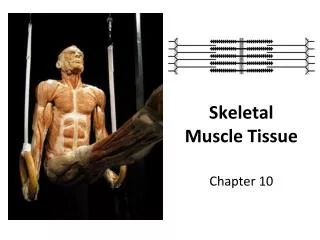
9 Skeletal Muscle Tissue
1.3k likes | 1.83k Vues
9 Skeletal Muscle Tissue. Section 1: Functional Anatomy of Skeletal Muscle Tissue. Learning Outcomes 9.1 Describe the organization of skeletal muscle at the tissue level. 9.2 Identify the structural components of a sarcomere.

9 Skeletal Muscle Tissue
E N D
Presentation Transcript
9 Skeletal Muscle Tissue
Section 1: Functional Anatomy of Skeletal Muscle Tissue Learning Outcomes 9.1 Describe the organization of skeletal muscle at the tissue level. 9.2 Identify the structural components of a sarcomere. 9.3 Describe the structural components of a thin filament and a thick filament. 9.4 Identify the components of the neuromuscular junction, and summarize the events involved in the neural control of skeletal muscles. 9.5 Describe the role of ATP in a muscle contraction, and explain the steps involved in the contraction of a skeletal muscle fiber.
Section 1: Functional Anatomy of Skeletal Muscle Tissue Muscle tissue One of the four primary tissue types Consists chiefly of muscle cells specialized for contraction Three types Cardiac In heart propelling blood through blood vessels Smooth Move fluids and solids along digestive tract Regulate diameters of small arteries Other functions
Section 1: Functional Anatomy of Skeletal Muscle Tissue Muscle tissue (continued) Three types (continued) Skeletal Move body by pulling on bones Each cell is a single muscle fiber Each muscle is an organ Primarily muscle cells plus connective tissues, nerves, and blood vessels
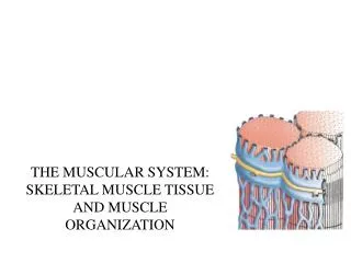
Figure 9 Section 1 Skeletal Muscle Tissue Skeletal muscle tissue contractions move the body by pulling on bones of the skeleton, making it possible for us to walk, dance, bite an apple, or play the ukulele. Cardiac Muscle Tissue Cardiac muscle tissue contractions in the heart propel blood through the blood vessels. Smooth Muscle Tissue Smooth muscle tissue contractions move fluids and solids along the digestive tract and regulate the diameters of small arteries, among other functions.
Section 1: Functional Anatomy of Skeletal Muscle Tissue Skeletal muscle tissue functions Produce skeletal movements Pull tendons and move bones Maintain posture and body position Skeletal muscle tension maintains body posture Support soft tissues Support weight of visceral organs and shield internal tissues from injury
Section 1: Functional Anatomy of Skeletal Muscle Tissue Skeletal muscle tissue functions (continued) Guard entrances and exits Openings of digestive and urinary tracts encircled by skeletal muscle (sphincters) Provide voluntary control of swallowing, defecation, and urination Maintain body temperature Some energy used for muscle contraction is released as heat Provide nutrient reserves Amino acids from muscle fibers can be released into circulation and used to produce glucose and energy
Module 9.1: Skeletal muscle anatomy Skeletal muscle Complex organ containing: Skeletal muscle fibers (contraction) Connective tissues (harness contractile forces) Blood vessels (nourish muscle fibers) Nerves (control contractions)
Module 9.1: Skeletal muscle anatomy Skeletal muscle connective tissues Tendon Bundle of collagen fibers that attach muscle to bone Collagen fibers extend into bone matrix providing firm attachment Also occurs as a sheet (= aponeurosis) Epimysium (epi-, on + mys, muscle) Dense layer of collagen fibers surrounding entire muscle Separates muscle from surrounding tissues and organs Connected to deep fascia
Module 9.1: Skeletal muscle anatomy Skeletal muscle connective tissues (continued) Perimysium (peri-, around) Fibrous layer that divides muscle into compartments or bundles of cells (= fascicles) Contains collagen and elastin fibers, blood vessels, and nerves Endomysium (endo-, inside) Surrounds individual muscle cells or fibers Loosely interconnects adjacent muscle fibers Contains capillaries, myosatellite (stem) cells, and axons of neurons that control muscle fibers
Module 9.1: Skeletal muscle anatomy Animation: Muscle Physiology: Muscle Layers Animation: Anatomy of Skeletal Muscles
Module 9.1: Skeletal muscle anatomy Skeletal muscle cell development Myoblasts (myo-, muscle + blastos, formative cell) fuse, forming multinucleate cells Develop into skeletal muscle fibers Each skeletal muscle fiber nucleus represents a myoblast Not all myoblasts fuse into developing muscle fibers Some remain in endomysium and help muscle repair
Figure 9.1 4 – 5 The development of a skeletal muscle fiber from myoblast to maturity Myoblasts Muscle fibers develop through the fusion of embryonic mesodermal cells called myoblasts. Myosatellite cell Over time, most of the myoblasts fuse together to form larger multinucleate cells. However, a few myoblasts remain within the tissue as myosatellite cells, even in adults. Nuclei Immature muscle fiber The multinucleate cells begin differentiating into skeletal muscle fibers as they enlarge and begin producing the proteins involved in muscle contraction. Myosatellite cell Mature skeletal muscle fiber Up to 30 cm in length
Module 9.1: Skeletal muscle anatomy Mature muscle cell characteristics Very large cells Can have diameter of 100 µm and length of 30 cm (12 in.) Each contains hundreds of nuclei just internal to plasma membrane (sarcolemma) (sarkos, flesh + lemma, husk) Genes in nuclei control production of enzymes and structural proteins for contraction Many nuclei = many genes = faster production Cytoplasm = sarcoplasm
Figure 9.1 5 – 6 A mature skeletal muscle fiber Myosatellite cell Mature skeletal muscle fiber Up to 30 cm in length Myofibrils Nuclei Sarcoplasm Mitochondria Sarcolemma
Module 9.1 Review a. Define tendon and aponeurosis. b. Describe the connective tissue layers associated with skeletal muscle tissue. c. How would severing the tendon attached to a muscle affect the muscle’s ability to move a body part?
Module 9.2: Skeletal muscle fiber anatomy Myofibrils Cylindrical structures 1–2 µm in diameter and as long as muscle fiber Hundreds to thousands comprise an individual muscle cell Each is banded and gives the skeletal muscle cells their banded appearance Made of protein filaments (= myofilaments) Thin filaments (primarily actin) Thick filaments (primarily myosin)
Figure 9.2 1 The myofibril, the source of a muscle fiber’s striations Myofibril Nuclei Sarcolemma Sarcoplasm Skeletal muscle fiber
Figure 9.2 2 A section of a muscle fiber, revealing its myofibrils, each of which is composed of myofilaments Sarcolemma Myofibril Thin filament Thick filament Mitochondria
Module 9.2: Skeletal muscle fiber anatomy Myofilament structure Have repeating functional units called sarcomeres (sarkos, flesh + meros, part) Approximately 10,000 sarcomeres/myofibril Each sarcomere has a 2-µm resting length Zone of overlap Thin and thick filaments interspersed Z lines Boundary of adjacent sarcomeres Interconnect thin filaments from adjacent sarcomeres Consist of proteins (actinins)
Module 9.2: Skeletal muscle fiber anatomy Myofilament structure (continued) A band Dense sarcomere region containing thick filaments I band Contains thin filaments (no thick) Extends from A band of one sarcomere to the next A band M line Connects central portion of each thick filament H band Lighter region around M line Contains only thick filaments (no thin)
Figure 9.2 3 Sarcomeres, the repeating functional units of myofilaments Arrangement of filaments in zone of overlap I band A band Myofibril M line Z line Sarcomere H band
Module 9.2: Skeletal muscle fiber anatomy Specialized parts of skeletal muscle cells Sarcolemma Separates sarcoplasm from interstitial fluid Maintains distribution of positive and negative charges on either side = Transmembrane potential All cells have a characteristic transmembrane potential Large changes in skeletal muscle transmembrane potential lead to contraction Are transmitted along entire muscle cell surface
Module 9.2: Skeletal muscle fiber anatomy Specialized parts of skeletal muscle cells (continued) Transverse tubules (T tubules) Narrow tubes from sarcolemma extending into sarcoplasm Transmembrane potential changes travel along T tubules to cell interior Encircle each sarcomere
Module 9.2: Skeletal muscle fiber anatomy Specialized parts of skeletal muscle cells (continued) Sarcoplasmic reticulum (SR) Similar to smooth endoplasmic reticulum of other cells Forms tubular network around each myofibril On either side of T tubule, forms expanded chambers (terminal cisternae) T tubule + terminal cisternae = triad
Figure 9.2 4 The transverse tubules and the sarcoplasmic reticulum T tubule encircling sarcomere at zone of overlap Transverse tubules (T tubules) Sarcolemma Transverse tubule Terminal cisternae Triad Position of M line
Module 9.2: Skeletal muscle fiber anatomy Specialized parts of skeletal muscle cells (continued) Sarcoplasmic reticulum (SR) (continued) Contains pumps moving calcium from sarcoplasm to SR SR calcium occurs as free ions and bound to proteins SR calcium concentrations can be 40,000× that of sarcoplasm Muscle contraction begins with SR calcium release
Figure 9.2 5 Sarcoplasmic reticulum (SR) Ca2+ Gated calcium channel (closed) Calcium ion pump Sarcoplasm The membrane of the sarcoplasmic reticulum, which contains calcium ion pumps
Module 9.2 Review a. Define transverse tubules. b. Describe the structural components of a sarcomere. c. Where would you expect the greatest concentration of Ca2+ to be in a resting skeletal muscle?
Module 9.3: Thick and thin filaments Thin filaments Attached to Z lines with actinin 5–6 µm in diameter, 1 µm in length Primarily actin Individual G-actin molecules (with active site for binding myosin) link together to form F-actin (filamentous) F-actin strand held together with nebulin
Module 9.3: Thick and thin filaments Thin filaments (continued) Also contain two regulatory proteins Tropomyosin Covers G-actin active sites and prevents actin–myosin interaction Attached to troponin Troponin Consists of three subunits Binds to tropomyosin (forms complex) Binds to G-actin (maintains position on actin) Binds two calcium ions (for activation during contraction) Animation: Muscle Physiology: Troponin
Figure 9.3 1 A longitudinal section of a sarcomere Myofibril Z line Thin filament Thick filament Actinin Z line The structure of thin filaments The attachment of thin filaments to the Z line at either end of a sarcomere Active site F-actin Troponin Nebulin Tropomyosin G-actin A thin filament, which is primarily composed of actin associated with other interacting proteins
Module 9.3: Thick and thin filaments Thick filaments 10–12 nm in diameter and 1.6 µm long Have core of titin Connect to Z lines Are elastic and recoil after stretching Contain ~300 myosin molecules
Module 9.3: Thick and thin filaments Thick filaments (continued) Myosin molecule Has long tail bound to other myosin molecules Has two globular subunits (= free head) Forms cross-bridges with actin during contraction Connection between head and tail allows a hinge-like (pivot) motion Animation: Muscle Physiology: Myosin Parts Animation: Muscle Physiology: Muscle Proteins
Module 9.3: Thick and thin filaments Sliding filament theory When muscles contract, thin filaments slide past thick filaments, and H bands and I bands get smaller Zones of overlap get larger Z lines approach each other A bands remain constant Sliding occurs in all sarcomeres of a myofibril Myofibril gets shorter Muscle cell gets shorter Animation: Muscle Physiology: Sarcromeres
Figure 9.3 3 The sliding filament theory I band A band A band I band H band Z line H band Z line Z line Z line Sarcomere contraction and filament sliding Sarcomere at rest
Module 9.3 Review a. Describe the components of thin filaments and thick filaments. b. What gives skeletal muscle its striated appearance? c. Briefly describe the sliding filament theory.
Module 9.4: Neuromuscular junction Neuromuscular junction (NMJ) Intercellular connection between motor neuron and skeletal muscle fiber Muscle fiber contracts only under control from the NMJ Only one NMJ per muscle fiber Although one motor neuron axon may branch to control multiple muscle cells A&P Flix: Events at the Neuromuscular Junction
Figure 9.4 1 Motor end plate Synaptic terminal Sarcoplasmic reticulum Myofibril Motor neuron Path of electrical impulse (action potential) Axon Neuromuscular junction Muscle fiber Myofibril Motor end plate The structural relationship between a skeletal muscle fiber and its lone neuromuscular junction
Module 9.4: Neuromuscular junction Neuromuscular junction (NMJ) (continued) Consists of: Synaptic terminal of neuron Has vesicles filled with neurotransmitter (acetylcholine [ACh]) Changes permeability of sarcolemma Motor end plate of muscle fiber Has junctional folds (creases) Contains acetylcholinesterase (AChE), an enzyme that breaks down ACh Synaptic cleft (space between neuron and muscle fiber) Also contains AChE
Figure 9.4 2 Vesicles containing ACh (red) Synaptic cleft Motor end plate AChE Junctional fold The locations of ACh and AChE in a resting neuro-muscular junction
Module 9.4: Neuromuscular junction Activities at the neuromuscular junction Electrical impulse (action potential) at the synaptic terminal causes vesicles of ACh to fuse with neuron plasma membrane = Exocytosis of ACh ACh diffuses across synaptic cleft and binds to receptors in motor end plate ACh binding allows Na+ to diffuse into the cell Sarcolemma generates action potential AChE inactivates receptors by quickly removing ACh from synaptic cleft
Figure 9.4 3 Arriving action potential Junctional fold The arrival of an action potential, the stimulus for ACh release
Figure 9.4 4 Sarcolemma of motor end plate Exocytosis of ACh into the synaptic cleft in response to arriving action potential
Figure 9.4 5 Na+ Na+ Na+ ACh receptor site Diffusion of ACh molecules and their binding to receptors on the motor end plate
Figure 9.4 6 Action potential AChE Generation of an action potential by the sudden inrush of sodium ions, and the breakdown of ACh by AChE
Module 9.4: Neuromuscular junction Action potential in the muscle cell Action potential (AP) generated at motor end plate sweeps across sarcolemma Effect is almost immediate since AP is an electrical event Event is brief since ACh has been removed and no other stimulus occurs until another AP at motor end plate Action potential sweeps down T tubules and causes calcium to be released from SR to sarcomeres causing muscle contraction = Excitation-contraction coupling Animation: Muscle Fiber Contraction
Figure 9.4 8 T tubule Sarcoplasmic reticulum (SR) Sarcoplasm Ca2+ Ca2+ Excitation-contraction coupling, the dumping of calcium ions onto sarcomeres as a result of the movement of an action potential down the T tubule