Download
1 / 51
520 likes | 708 Vues
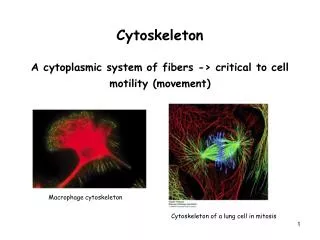
Cytoskeleton A cytoplasmic system of fibers -> critical to cell motility (movement). Macrophage cytoskeleton. Cytoskeleton of a lung cell in mitosis. The cytosol: 20-30 w% of cytosol are proteins -> ¼ - ½ of total protein is in cytosol Protein conc. 200-400 mg/ml -> complexes of protein

E N D
CytoskeletonA cytoplasmic system of fibers -> critical to cell motility (movement) Macrophage cytoskeleton Cytoskeleton of a lung cell in mitosis
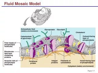
The cytosol: 20-30 w% of cytosol are proteins -> ¼ - ½ of total protein is in cytosol Protein conc. 200-400 mg/ml -> complexes of protein It is believed that cytosol is highly organized -> Most soluble proteins are - bound to filaments - localized in specific regions
Eukaryotes Bacteria Actin filaments (AF) Intermediate filaments (IF) microtubule (MT)
Filament network in the cell Filament network (fluorescence) From the nucleus to the plasma membrane Microtubules network Starting from the MT center near nucleus
Cytoskeleton supporting the plasma membrane in human red blood cells
1. Microfilaments and Actin Structures Actin cytoskelet in a moving cell
1. Microfilaments and Actin Structures Actin monomers assemble into long helical polymers with polarity
Actin Filaments grow faster at (+) end than at (-) end The rate of addition of ATP-G-actin is much faster at the (+) end than at the (-) end (rate of dissociation is similar) -> lower critical concentration (Cc) at (+) end in steady state -> filament grows preferentially at the (+) end If actin conc. is between Cc- and Cc+ (steady state) -> actin subunits flow through filaments by attaching to (+) end and dissociating from (-) end Treadmilling phenomen -> involved in movement of cells
Capping Proteins Block Assembly and Disassembly at Actin Filament Ends The presence of these 2 proteins at opposite ends prevent actin from dissociating during muscle contraction
Actin Filament Branching Nucleation of branching mediated by Arp2/3
Movement of invaders inside the cell Most infections are spread by lysed cells. Some Bacteria (Listeria monocytogenes) or viruses (vaccinia – related to smallbox virus) escape from cell on the end of a polymerizing actin filament. These organisms or viruses move through the cytosol at rates of 11μm/min. Actin generates the force necessary for movement
Proteins that organize Microfilaments into networks Forms bundles Filamin forms networks
Filaments attached to Membranes Microvilli on an epithelial cell showing polarity of actin filaments
Myosins - Cellular Motor Proteins Tail: -> Locatized to cellular membranes -> vesicle attached (cargo) Form thick filaments in muscles S1 motor domain Head -> Motor domain (S1) -> ATP depentend
Myosin heads walk along actin filaments -> towards (+) end Sliding-filament assay: Myosin tail absorbed onto glass surface -> a solution of actin filaments allowed to flow through In presence of ATP myosin heads walk towards (+) end of actin filaments -> sliding of filaments -> Movement of labeled actin filaments
Myosins – Motor proteins responsible for cell movement These are the most important 3 myosins (out of ca. 40 we have in humans) Loss of more specialized ones -> causes deafness/blindness
Myosin motion along actin Length of the neck domain -> determines rate of movement Step size -> Moves in 72 nm steps
Skeletal muscle contraction is regulated by Calcium and actin binding proteins Tropomyosin (TM) Troponin (TN)
Cell Locomotion Coordination of motions generated by different parts of the cell Movement of fish epidermal cell Cell locomotion mechanism: Includes actin polymerization and branching-generated movement at the edge, assembly of adhesion structures, and contractions mediated by myosin II
Myosin V Carries Many Cargoes Myosin V: -> carries secretory vesicles, organelles,... -> Used to prepare nucleus for mitosis -> used to segregate organelles
2. Intermediate Filaments • IF differ in stability, size and structure from other cytoskeleton fibers: • - IF are extremely stable (hair, nails, wool) • 10 nm diameter • α-helical rods -> assemble into ropelike filaments • assemble from different IF proteins • assembly through several intermediate structures Keratin and lamin IF Intermediate structures in the assembly of IF
Cross-links between Microtubules and Intermediate Filaments in Fibrioblast cells Microtubules (red), Intermediate Filaments (blue), connection between fibers (green)
3. Microtubules Kinesin-powered movement of a vesicle along a microtubule • Microtubules are involved in cell movement: • Beating of cilia and flagella • transport of vesicles in the cytoplasm Microtubules organizing center (MTOC)
Microtubules organization • 2 type of MT in cells: • Stable and long-lived (found in non-replication cells) -> in cilia, flagella, neurons • unstable and short-lived (found in mitosis) -> spindle-shaped apparatus that partitions chromosomes equally to daughter cells
Microtubules Arrangement Flagella
Microtubules assemble from Microtubule Organizing Centers (MTOCs) MTOCs in non-mitotic cells -> centrosomes
Microtubules Assembly preferably at (+) end Nucleation of microtubule assembly -> Treadmilling
Microtubules Assembly/Disassembly Colchicine and Taxol: Drugs that interfere with Microtubules Assembly/Disassambly Colchicine: 2500 years ago Egyptians treated heart problems Nowadys: treatment of gout, skin and joint diseases Taxol: (stabilizes Microtubules) Anticancer agents -> treatment of ovarian cancer
Microtubules Dynamic Instability Presence of GTP-β-tubulin cap determines stability
Kinesin and Dynein – two Families of Motor Proteins Responsible for Transport along Microtubules Microtubules based vesicle transport
Kinesin-catalysed Vesicle Transport Carries cargo
Kinesin-1 uses ATP to walk down a microtubule to the (+) end
Dynein-catalysed Vesicle Transport Moves towards the (-) end of microtubules
Microtubule motors in a Cell Kinesins -> transport to cell periphery (+) Dyneins -> transport to cell center (-)
Cooperation of Myosin and Kinesin at the cell cortex Secretory vesicles are handed over from Kinesin to Myosin -> last part of secretory pathway
Rotory Motors – Cilia and Flagellar Bacterial Flagella (E. coli, Salmonella) Rotation of Flagellar -> Motion Sperm
Motion of E. coli The points show the locations of the bacterium at 80 ms intervals. Changing of direction: Tumbling is caused by an abrupt reversal of the fagellar motor A second reversal restores smooth swimming -> almost always in a different direction
Reversal of the direction of Flagellar Rotation is obtained by Proton Transport through Motor Proton flow drives flagella rotation !!!