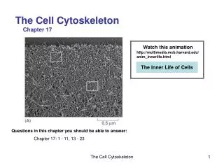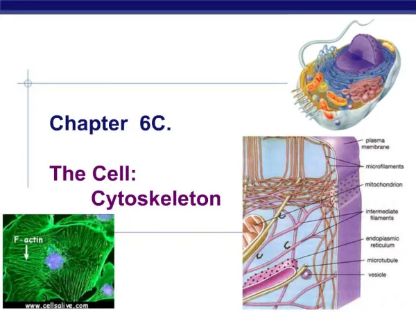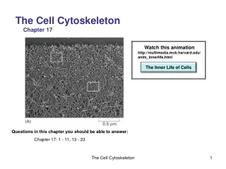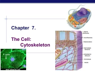
CHAPTER 16 THE CYTOSKELETON
CHAPTER 16 THE CYTOSKELETON. THE SELF-ASSEMBLY AND DYNAMIC STRUCTURE OF CYTOSKELETAL FILAMENTS HOW CELLS REGULATE THEIR CYTOSKELETAL FILAMENTS MOLECULAR MOTORS THE CYTOSKELETON AND CELL BEHAVIOR. THE SELF-ASSEMBLY AND DYNAMIC STRUCTURE OF CYTOSKELETAL FILAMENTS.

CHAPTER 16 THE CYTOSKELETON
E N D
Presentation Transcript
CHAPTER 16 THE CYTOSKELETON • THE SELF-ASSEMBLY AND DYNAMIC STRUCTURE OF CYTOSKELETAL FILAMENTS • HOW CELLS REGULATE THEIR CYTOSKELETALFILAMENTS • MOLECULAR MOTORS • THE CYTOSKELETON AND CELL BEHAVIOR
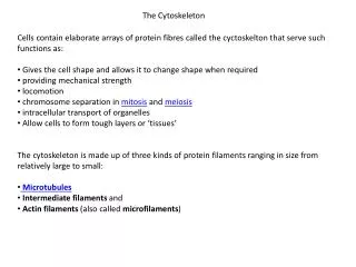
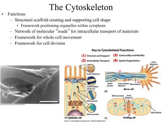
THE SELF-ASSEMBLY AND DYNAMIC STRUCTURE OF CYTOSKELETAL FILAMENTS • Three Types of Cytoskeletal Filaments • Nucleation Is the Rate-limiting Step in the Formation of a Cytoskeletal Polymer • The Two Ends of a Microtubule and of an Actin Filament Are Distinct and Grow at Different Rates • Filament Treadmilling and Dynamic Instability Are Consequences of Nucleotide Hydrolysis by Tubulin and Actin • Intermediate Filaments Impart Mechanical Stability to Animal Cells • Filament Polymerization Can Be Altered by Drugs
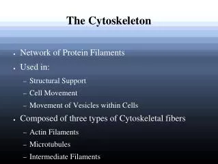
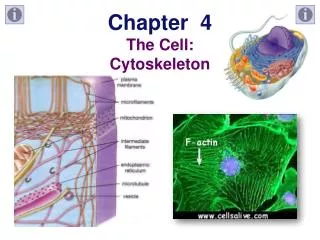
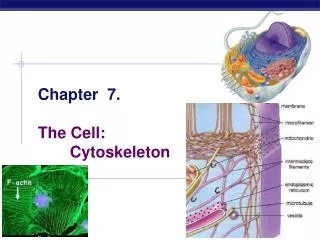
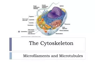
Three Types of Cytoskeletal Filaments • Actin filaments (5-9 nm diameter) • Actin subunits • Locomotion, muscle contraction • Intermediate filaments (10 nm diameter) • Various coiled coil protein subunits • (lamins, vimentin, keratin) • Structural roles • Microtubules (25 nm diameter) • Tubulin subunits • Intracellular transport
Nucleation Is the Rate-limiting Step in the Formation of a Cytoskeletal Polymer
The Two Ends of Microtubules and Actin Filaments Are Distinct and Grow at Different Rates
Filament Treadmilling and Dynamic Instability Are Consequences of Nucleotide Hydrolysis
Intermediate Filaments Impart Mechanical Stability to Animal Cells
Filament Polymerization Can Be Altered by Drugs • TABLE 16–2 Drugs That Affect Actin Filaments and Microtubules • ACTIN-SPECIFIC DRUGS • Phalloidin binds and stabilizes filaments • Cytochalasin caps filament plus ends • Swinholide severs filaments • Latrunculin binds subunits and prevents their polymerization • MICROTUBULE-SPECIFIC DRUGS • Taxol binds and stabilizes microtubules • Colchicine, colcemid binds subunits and prevents their polymerization • Vinblastine, vincristine binds subunits and prevents their polymerization • Nocodazole binds subunits and prevents their polymerization
HOW CELLS REGULATE THEIR CYTOSKELETALFILAMENTS • Microtubules Are Nucleated by a Protein Complex Containing g-tubulin in the Centrosomes of Animal Cells • Regulatory Proteins Bind to Free Subunits, Filaments Sides and Filament Ends • Extracellular Signals Can Induce Major Cytoskeletal Rearrangements
Microtubules Are Nucleated by a Protein Complex Containing g-tubulin in the Centrosomes of Animal Cells
Regulatory Proteins Bind to Free Subunits, Filaments Sides and Filament Ends
Extracellular Signals Can Induce Major Cytoskeletal Rearrangements
MOLECULAR MOTORS • Actin-based Motor Proteins Are Members of the Myosin Superfamily • There Are Two Types of Microtubule Motor Proteins: Kinesins and Dyneins • Motor Proteins Generate Force by Coupling ATP Hydrolysis to Conformational Changes • Cilia and Flagella Are Motile Structures Built from Microtubules and Dyneins
Actin-based Motor Proteins Are Members of the Myosin Superfamily • Myosin II is the muscle motor • Other myosins have other functions
There Are Two Types of Microtubule Motor Proteins: Kinesins and Dyneins
Motor Proteins Generate Force by Coupling ATP Hydrolysis to Conformational Changes
Cilia and Flagella Are Motile Structures Built from Microtubules and Dyneins
THE CYTOSKELETON AND CELL BEHAVIOR • Mechanisms of Cell Polarization Can Be Readily Analyzed in Yeast Cells • Directional Assembly Dictates the Direction of Cell Migration • The Complex Morphological Specialization of Neurons Depends on The Cytoskeleton
Mechanisms of Cell Polarization Can Be Readily Analyzed in Yeast Cells
Directional Assembly Dictates the Direction of Cell Migration
The Complex Morphological Specialization of Neurons Depends on The Cytoskeleton
CHAPTER 18 THE MECHANICS OF CELL DIVISION • AN OVERVIEW OF M PHASE • MITOSIS • CYTOKINESIS
AN OVERVIEW OF M PHASE • Cohesins and Condensins Help Configure Replicated Chromosomes for Segregation • The Cytoskeleton Carries Out Both Mitosis and Cytokinesis • Two Mechanisms Help Ensure That Mitosis Always Precedes Cytokinesis • M Phase Depends on DNA Replication and Centrosome Duplication in the Preceding Interphase • M Phase Is Traditionally Divided into Six Stages
Cohesins and Condensins Help Configure Replicated Chromosomes for Segregation
Two Mechanisms Help Ensure That Mitosis Always Precedes Cytokinesis • 1. Proteins required for cytokinesis are inactivated by M-Cdk during mitosis • 2. The remnants of the mitotic spindle are required for assembly of the contractile ring before cytokinesis
M Phase Depends on DNA Replication and Centrosome Duplication in the Preceding Interphase
MITOSIS • Microtubule Instability Increases Greatly at M Phase • Interactions Between Opposing Motor Proteins and Microtubules of Opposite Polarity Drive Spindle Assembly • Kinetochores Attach Chromosomes to the Mitotic Spindle • Anaphase Is Delayed Until All Chromosomes Are Positioned at the Metaphase Plate • Sister Chromatids Separate Suddenly at Anaphase • Kinetochore Microtubules Disassemble at Both Ends During Anaphase A • Both Pushing and Pulling Forces Contribute to Anaphase B • At Telophase, the Nuclear Envelope Re-forms Around Individual Chromosomes
Microtubule Instability Increases Greatly at M Phase • MAPs stabilize • Catastrophins destabilize • Rapid turnover results in survival of only productive (capped, attached, stabilized) microtubules
Interactions Between Opposing Motor Proteins and Microtubules of Opposite Polarity Drive Spindle Assembly • (-) end motors (like Kar3p) organize tubules at spindle poles • (+) end motors (like Cin8p) push tubules of opposite orientation against each other
CYTOKINESIS • The Microtubules of the Mitotic Spindle Determine the Plane of Animal Cell Division • Actin and Myosin II in the Contractile Ring Generate the Force for Cytokinesis • Membrane-enclosed Organelles Must Be Distributed to Daughter Cells During Cytokinesis • Mitosis Can Occur Without Cytokinesis • The Phragmoplast Guides Cytokinesis in Higher Plants • The Elaborate M Phase of Higher Organisms Evolved Gradually from Procaryotic Fission Mechanisms
The Microtubules of the Mitotic Spindle Determine the Plane of Animal Cell Division
Actin and Myosin II in the Contractile Ring Generate the Force for Cytokinesis
Bacterial Fission - a model for M phase& FtsZ - a bacterial tubulin homolog