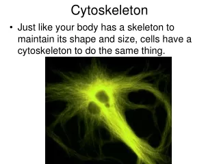
Cytoskeleton
Cytoskeleton. Cristina Botello-Godoy, Karina Garcia, Takara Reed, Aida Rogonich. Animals, fungi, protozoans, algae, etc. Much larger & more complex, as are the cells Structure’s include: shape & strong cell wall, allow for cell movement (within cells & endocytosis), cell division. Bacteria


Cytoskeleton
E N D
Presentation Transcript
Cytoskeleton Cristina Botello-Godoy, Karina Garcia, Takara Reed, Aida Rogonich
Animals, fungi, protozoans, algae, etc. Much larger & more complex, as are the cells Structure’s include: shape & strong cell wall, allow for cell movement (within cells & endocytosis), cell division Bacteria Smaller & less complex, as are the cells Structure’s include: shape & strong cell wall, cell division Eukaryotic vs. Prokaryotic Cytoskeleton
Prokaryotic Cytoskeleton • Recent study has found that bacteria cells too consist of a cytoskeleton • Research is based on the fact that the proteins of prokaryotic & eukaryotic cells are homologues • Evolution is diverse between the two cell types, so more concepts allowed for this approach
Prokaryotic Cytoskeleton • Network of structural filaments • 3-D structure • Maintains cell shape, structure, protection, polarity, and cell division • Some structures are still UNIDENTIFIED • Proteins similar to those of eukaryotic cells discovered
FtsZ: tubulin-like forms filaments, but they do not group into tubules maintains organization required for cell division MreB: actin-like determines cell shape positions polar proteins forms a network (helix shaped) under the cytoplasmic membrane, that directs proteins Protein Functions
CreS: crescentin intermediate filament like maintains cell shape and form of bacteria filament connects from pole to pole ParM: actin-like allow for cell division acts like kinetochore complez separation of plasmids by the filaments is analogous to microtubules allowing cell division during eukaryotic mitosis Protein Functions
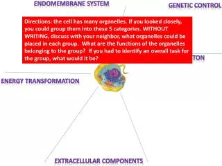
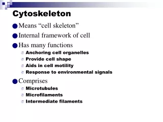
Cytoskeleton Function • The Cytoskeleton has many important functions. One of its most important functions is to provide structure for the cell, it makes sense if you break up the word cyto-skeleton. • -The cytoskeleton also acts as a scaffolding, with this the organelles in the cell are able to stay attached. • -The cytoskeleton is also used for the movement of the cell. The internal movement of cell organelles, as well as cell locomotion and muscle fiber contraction could not take place if it wasn’t for the cytoskeleton. • -The cytoskeleton is also required for the division of cells during mitosis or meiosis.
Endoplasmic Reticulum (E.R.) • The Endoplasmic Reticulum is the factory in the cell that produces proteins and lipids of most of the cells’ s organelles. • The E.R is mainly responsible for transporting proteins and other carbohydrates to wherever they are needed in the cell. • The E.R is composed of many folds, say you fold a piece of gum over and over that’s how the E.R looks. • There are two types of E.R, a Rough E.R and a Smooth E.R • The Rough E.R is rough because it has ribosome attached to it. In the Rough E.R proteins are made . • The Smooth E.R doesn’t have any thing attached to it, that’s why its called smooth • The Smooth E.R is the one that releases the proteins produced in the Rough E.R
Cell Shape • The cell shape depends on what type of cell it is • If the cell is an animal cell than most likely the cell is circular because it doesn’t contain a cell wall • If the cell is rectangular or like a square then the cell is an animal cell, the cell takes the shape of the cell wall
Cytoskeleton Structure • Imagine walking into George’s basement. All you see is a bunch of glow in the dark cables hanging over your head. These cables surround the machines that operate our school. These cables are the cytoskeleton, which surrounds the cells that help function our body. =)
Cytoskeleton Structure • The Cytoskeleton is a complex three-dimensional structure that is found in the cytoplasm of the cell. It’s composed of fibers primary microtubules being the thickest of three, microfilaments, the thinnest, and intermediate filaments in the middle range.
Microtubules are one of the components of the cytoskeleton. They are rigid hollow rods that have a diameter of 25 nanometers and length of anywhere between 200 nanometers and 25 micrometers. Microtubules serve as structural components within cells and are involved in many cellular processes including mitosis, cytokinesis, and vesicular transport. Microtubules
Centrosomes organize the microtubules. They also regulate cell-cycle progression. This occurs in the cytoplasm attached to the outside of the nucleus. In nine triplet sets (star shaped), microtubules form Centrioles surrounded by a solid mass of protein called the “pericentriolar material.” In nine doublets oriented about additional microtubules (wheel-shaped) microtubules form cilia & flagella. Centrosomes, Centrioles, Cilia & Flagella
ANIMAL The centrosome, also called the "microtubule organizing center“ (MTOC), is a region in the cell where microtubles are formed. Within an animal cell centrosome, there is a pair of small organelles known as the centrioles which each make up of a ring of nine groups of microtubules. (see picture). CELLS: The two centrioles are arranged so that one is perpendicular to the other. During animal cell division, the centrosome divides and the centrioles replicate which results in two centrosomes that each have its own pair of centrioles. The two centrosomes move to opposite ends of the nucleus, and from each centrosome, microtubules grow into a "spindle" which is responsible for separating replicated chromosomes into the two daughter cells (Mitosis process). Centrosomes & Centrioles
Centrosomes & Centrioles • PLANT CELLS: • Plant cells have centrosomes that function much like animal cell centrosomes. However, unlike centrosomes in animal cells, they do not have centrioles.
Both cilia and flagella are created from microtubules, and both supply: “Locomotion” for the cells (like sperm) And the movement of fluid past the cells Both have the same basic structure. If the microtubules are short, they are cilia Or only one or a few long ones, they are flagella. Each cilium or flagellum is made of: a cylindrical collection of 9 evenly-spaced microtubules, each with an incomplete microtubule attached to it. This gives the structure a "figure 8" appearance when view in cross section. (see bottom picture). 2 single microtubules run up through the center of the bundle, completing the so-called "9+2" pattern. The entire assembly is enclosed in a membrane that is basically an expansion of the plasma membrane. Cilia & Flagella
Solid rods about 7 nm in diameter made from actin a globular protein, in the form of a double twisted chain Structural role is to bear tension, resist the pulling forces within the cell Microfilaments (actins filaments) Actin subunit 7 nm
Forms a 3-D network called the cortex just inside the plasma membrane to help support the cell’s shape make up the core of microvilli of intestinal cells Microfilaments (actins filaments)
Microfilaments (actins filaments) • Microfilaments are known to assist in cell motility. • In muscles cells thousands of actin filaments are arranged parallel to one another along the length of a muscle cell, interlinking with thicker filaments made of the protein myosin. Myosin acts as a motor protein as projections walk along the actin filaments.
Muscle cell Actin filament Myosin filament Myosin arm (a) Myosin motors in muscle cell contraction
Microfilaments (actins filaments) Microfilaments (actins filaments) • Amoeboid movement interaction of actin filaments with myosin near the cell’s trailing end squeezes the interior fluid forward into the pseudopodium Cortex (outer cytoplasm): gel with actin network Extending pseudopodium Inner cytoplasm: sol with actin subunits
Microfilaments (actins filaments) • Main functions: • Maintenance of cell shape (tension-bearing elements) • Changes in the cell shape • Muscle contraction • Cell motility • Myosin • Pseudopodia • Cytoplasmic streaming
Intermediate Filaments Intermediate Filaments • Diameter of 8-12nm, • As well as Microfilaments, it is specialized for bearing tensions. • Constructed of different molecular subunits belonging to a family of proteins whose members include the keratin Keratin proteins Fibrous subunit (keratins coiled together)
Intermediate Filaments • Are more permanent fixtures of cells then microtubules and microfilaments • After cells die intermediate filaments are still found on the cells • Experiments suggest that intermediate filaments are of greater importance in reinforcing the shape of a cell and organelles . • Nucleolus is surrounded by intermediate filaments that hold it in place within the cell • Also makes up the nuclear lamina inside the nuclear envelope
Resources Internet: • http://student.ccbcmd.edu/courses/bio141/lecguide/unit3/eustruct/cytosk.html • http://en.wikipedia.org/wiki/Cytoskeleton • http://en.wikipedia.org/wiki/Prokaryotic_cytoskeleton • www.bio.miami.edu • www.cancerquest.org • www.darwin.nmsu.edu • http://www.cellsalive.com/cell/centriol.htm • http://users.rcn.com/jkrmball.ma.ultranet/BiologyPages/c/Cytoskeleton.htm • http://www.ncbI.nlm.nih.gov/books/bv.fcgi?indexed+google&rid+cooper.section1820 Books: • AP Biology (7th Edition) by Campbell Reece