Download
1 / 1
10 likes | 113 Vues
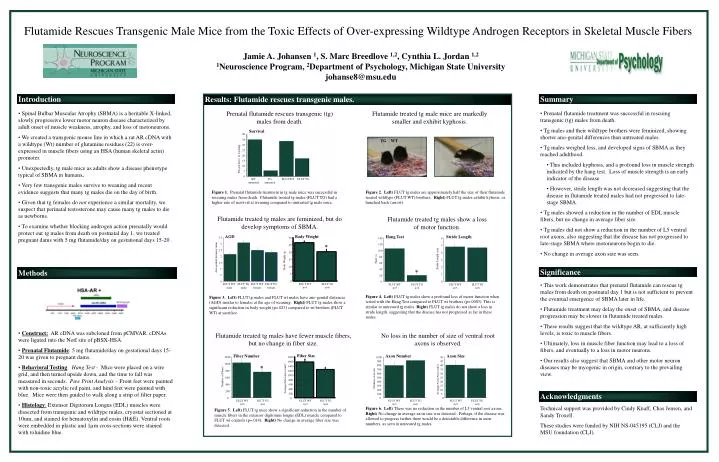
Introduction. Results: Flutamide rescues transgenic males. Summary. Spinal Bulbar Muscular Atrophy (SBMA) is a heritable X-linked, slowly progressive lower motor neuron disease characterized by adult onset of muscle weakness, atrophy, and loss of motoneurons.

E N D
Introduction Results: Flutamide rescues transgenic males. Summary • Spinal Bulbar Muscular Atrophy (SBMA) is a heritable X-linked, slowly progressive lower motor neuron disease characterized by adult onset of muscle weakness, atrophy, and loss of motoneurons. • We created a transgenic mouse line in which a rat AR cDNA with a wildtype (Wt) number of glutamine residues (22) is over-expressed in muscle fibers using an HSA (human skeletal actin) promoter. • Unexpectedly, tg male mice as adults show a disease phenotype typical of SBMA in humans. • Very few transgenic males survive to weaning and recent evidence suggests that many tg males die on the day of birth. • Given that tg females do not experience a similar mortality, we suspect that perinatal testosterone may cause many tg males to die as newborns. • To examine whether blocking androgen action prenatally would protect our tg males from death on postnatal day 1, we treated pregnant dams with 5 mg flutamide/day on gestational days 15-20 . Prenatal flutamide rescues transgenic (tg) males from death. Flutamide treated tg male mice are markedly smaller and exhibit kyphosis. • Prenatal flutamide treatment was successful in rescuing transgenic (tg) males from death. • Tg males and their wildtype brothers were feminized, showing shorter ano-genital differences than untreated males. • Tg males weighed less, and developed signs of SBMA as they reached adulthood. • This included kyphosis, and a profound loss in muscle strength indicated by the hang test. Loss of muscle strength is an early indicator of the disease. • However, stride length was not decreased suggesting that the disease in flutamide treated males had not progressed to late-stage SBMA. • Tg males showed a reduction in the number of EDL muscle fibers, but no change in average fiber size. • Tg males did not show a reduction in the number of L5 ventral root axons, also suggesting that the disease has not progressed to late-stage SBMA where motoneurons begin to die. • No change in average axon size was seen. Survival 40 TG WT 35 30 25 20 Percent alive at weaning 15 10 5 0 WT untreated TG untreated FLUT WT FLUT TG Figure 1. Prenatal flutamide treatment in tg male mice was successful in rescuing males from death. Flutamide treated tg males (FLUT TG) had a higher rate of survival at weaning compared to untreated tg male mice. Figure 2. Left) FLUT tg males are approximately half the size of their flutamide treated wildtype (FLUT WT) brothers. Right) FLUT tg males exhibit kyhosis, or hunched back (arrow). Flutamide treated tg males are feminized, but do develop symptoms of SBMA. Flutamide treated tg males show a loss of motor function. AGD Hang Test 3.5 140 Body Weight 30 3 120 Stride Length * 25 6 2.5 100 2 20 80 5 Ano-genital distance (mm) Time (s) 1.5 60 15 4 Body Weight (g) 1 Significance Methods 40 * Stride Length (cm) 10 3 .5 20 5 2 0 0 • This work demonstrates that prenatal flutamide can rescue tg males from death on postnatal day 1 but is not sufficient to prevent the eventual emergence of SBMA later in life. • Flutamide treatment may delay the onset of SBMA, and disease progression may be slower in flutamide treated males. • These results suggest that the wildtype AR, at sufficiently high levels, is toxic to muscle fibers. • Ultimately, loss in muscle fiber function may lead to a loss of fibers, and eventually to a loss in motor neurons. • Our results also suggest that SBMA and other motor neuron diseases may be myogenic in origin, contrary to the prevailing view. FLUT WT male FLUT TG male FLUT WT female FLUT TG female FLUT WT n=5 FLUT TG n=6 0 1 FLUT WT n=5 FLUT TG n=6 0 Figure 4. Left) FLUT tg males show a profound loss of motor function when tested with the Hang Test compared to FLUT wt brothers (p=.0003). This is similar to untreated tg males. Right) FLUT tg males do not show a loss in stride length, suggesting that the disease has not progressed as far in these males. Figure 3. Left) FLUT tg males and FLUT wt males have ano-genital distances (AGD) similar to females at the age of weaning. Right) FLUT tg males show a significant reduction in body weight (p=.023) compared to wt brothers (FLUT WT) at sacrifice. FLUT WT n=5 FLUT TG n=6 • Construct: AR cDNA was subcloned frompCMVAR. cDNAs were ligated into the NotI site of pBSX-HSA. • Prenatal Flutamide: 5 mg flutamide/day on gestational days 15-20 was given to pregnant dams. • Behavioral Testing: Hang Test - Mice were placed on a wire grid, and then turned upside down, and the time to fall was measured in seconds. Paw Print Analysis – Front feet were painted with non-toxic acrylic red paint, and hind feet were painted with blue. Mice were then guided to walk along a strip of filter paper. • Histology: Extensor Digitorum Longus (EDL) muscles were dissected from transgenic and wildtype males, cryostat sectioned at 10um, and stained for hematoxylin and eosin (H&E). Ventral roots were embedded in plastic and 1µm cross-sections were stained with toluidine blue. Flutamide treated tg males have fewer muscle fibers, but no change in fiber size. No loss in the number of size of ventral root axons is observed. Axon Size 50 Fiber Size Fiber Number Axon Number 45 1800 1200 1000 40 900 1600 1000 * 800 35 1400 30 700 800 1200 25 600 Average Axon Area (um2) 1000 Number of Fibers 20 600 500 Number of Axons Average Fiber Area (um2) 800 15 400 400 600 300 10 400 Acknowledgments 5 200 200 200 0 100 FLUT WT n=5 FLUT TG n=6 0 0 0 FLUT WT n=5 FLUT TG n=6 FLUT WT n=5 FLUT TG n=6 FLUT WT n=5 FLUT TG n=6 Technical support was provided by Cindy Knaff, Chas Jensen, and Sandy Troxell. These studies were funded by NIH NS-045195 (CLJ) and the MSU foundation (CLJ). Figure 6. Left) There was no reduction in the number of L5 ventral root axons. Right) No change in average axon size was detected. Perhaps, if the disease was allowed to progress farther there would be a detectable difference in axon numbers, as seen in untreated tg males. Figure 5. Left) FLUT tg mice show a significant reduction in the number of muscle fibers in the extensor digitorum longus (EDL) muscle compared to FLUT wt controls (p=.014). Right) No change in average fiber size was detected. Flutamide Rescues Transgenic Male Mice from the Toxic Effects of Over-expressing Wildtype Androgen Receptors in Skeletal Muscle Fibers Jamie A. Johansen 1, S. Marc Breedlove 1,2, Cynthia L. Jordan 1,2 1Neuroscience Program, 2Department of Psychology, Michigan State University johanse8@msu.edu