Open Pelvic Fracture
490 likes | 928 Vues
Open Pelvic Fracture. Intern 蕭福慶. Brief History. 42 y/o female, denied systemic disease Pedestrian hit by car on 94.12.9 Lower limbs numbness, back pain Open pelvic fracture, bilateral scapular fracture Operation in 和平 Hospital. Event 1. X-ray: L’t pelvic fracture

Open Pelvic Fracture
E N D
Presentation Transcript
Open Pelvic Fracture Intern 蕭福慶
Brief History • 42 y/o female, denied systemic disease • Pedestrian hit by car on 94.12.9 • Lower limbs numbness, back pain • Open pelvic fracture, bilateral scapular fracture • Operation in 和平 Hospital
Event 1 • X-ray: L’t pelvic fracture • Spine CT: no obvious abnormality • 12.11: hypotension, tachycardia • 12.12: fecal material in drain • Abd CT: gas at rectal wall • Laparotomy with sigmoid and colostomy
Question 1 • Does rectal injury have correlation with open pelvic fracture?
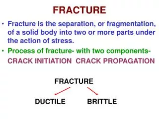
Open Pelvic Fracture • A direct communication between a skin, rectal, or vaginal wound and the fracture • Motality: 5% to 50% (25~30%)
Risk • Incidence of anorectal injury: 18~64% • Incidence of urogenital injury: 23~53% • Rectal injury: pubic symphysis, SI joint • Bladder injury: SI joint, pubic symphysis, fractures of the sacrum • Urethral injury: superior, inferior pubic rami, pubic symphysis
Examination • Anteroposterior pelvic X-ray • Inspection of the perianal tissues • Digital rectal examination • Sigmodoiscopy or proctoscopy, • Pelvic CT: localised extraluminal gas haemorrhage bowel wall thickening
Treatment • Pelvic fracture stabilization • Wound debridement • Selective diverting colostomy • Colostomy takedown (6weeks~3months)
Brief History • 12.12: Shock, dopamin was used (hypotension, respiratory failure, elevated liver function, oliguria, pancytopenia, limbs discoloration) • Doppler: no flow at bilateral dorsa pedis, medial, lateral malleolus arteries in ankles • 12.17: tentative fasciotomy
Event 2 • 94.12.22: CVS: PGE1, pletaal NS: suspect T8 injury • 12.23: Wound: Proteus mirabilis, Klebsiella pneumoniae, Pseudomonas aeruginosa • 12.27: Left AK amputation • 95.1.2: Right AK amputation
Question 2 • Why does her four limbs have gangrene change?
Symmetrical Peripheral Gangrene • Symmetrical distal ischemic damage in two or more sites • Absence of large vessel obstruction • Rare(?) • Motality: up to 40% with DIC • Amputation: 50% • Ischemia of other organ (gut)
Mechanism • Vasospasm • Pathology in microcirculation • Slugging of platelet or fibrin degeneration product
Risk • Disseminated intravascular coagulation • Sepsis (Streptococcus, Staphylococcus) • Vasopressor • Malignant disease(paraneoplastic syndrome) • Ergotism • Protein C deficiency • Cold injury • Scleroderma, polymyalgia rheumatica • Impaired renal function • Splenectomy • Diabetes mellitus • Immunosuppression • Alcoholism
Course • Marked coldness, pallor, cyanosis or pain in the extremity • Progress rapidly to acrocyanosis • Gangrene
Treatment • Control underlying problem (DIC, sepsis, vasopressor) • Local or intravenous infusion of an α-blocker (phentolamine, chlorpromazine) • Intravenous infusion of prostaglandin (epoprostenol) • Sympathetic blockade (ganglion block or intravenous trimethaphan therapy) • Intravenous nitropruside therapy • Topical nitroglycerine ointment • Amputation (usually not emergency)
Brief History • 12.12: Shock: oliguria • 12.14~: H/D • BUN/Cre: 127.6/5.6 • U/O: 250-300ml • Bilateral pleural effusion, pitting edema
Event 3 • 12.27: Left AK amputation • Contrast CT was need to evaluate the situation of infection (possible abscess)
Question 3 • Can we perform contrast CT on the patient with acute renal failure?
Contrast-induced Nephropathy • Within 48 hours after administration of contrast media. • Increased Serum creatinine > 44μmol/L (0.5 mg/dl) • Relative increase of at least 25% • Oliguria 2~5 days, recover on Day 7
Mechanism • Renal hemodynamic change: • medullar hypoxia • Direct toxic effect: • tubular epithelial cell • Osmolality: • compress intrarenal microcirculation • decreased glomerular filtration rate
Prevention • Hydration (U/O>150 mL/hr for the first 6 hours after the procedure) • IOCM, iso-osmolar contrast medium(Iodixanol) • Low contrast volume (<100 mL, Spacing at least 10 days) • N-acetylcysteine(NAC)(600 mg, by mouth, twice a day (two dosages before and two doses after contrast exposure)) • Hemofiltration for critical ill or ICU
Treatment • Supportive management • Hemodialysis (eGFR<15)
Take home message 1 • Open pelvic fracture: • High motality • Stabilize hemodynamics • Complete examination(rectal, vaginal, wound) • Colostomy, debridement
Take home message 2 • Symmetrical peripheral gangrene • Early notice of ischemic sign • Beware of multiple organ ischemia • Control DIC, sepsis • Decrease the use of vasopressor as possible • Amputation rate: 50%
Take home message 3 • Contrast-induced nephropathy • Evaluate risk • Alternative examination • Hydration if could tolerate • Low contrast volume • N-Acetylcysteine • Hemofiltration in critical ill
Thanks for your attention Thank you
Reference-1 • Bircher et al. Pelvic trauma management within the UK: a reflection of a failing trauma service; Injury, Int. J. Care Injured (2004) 35, 2—6 • Collinge et al. Soft tissue injuries associated with pelvic fractures; Orthop Clin N Am 35 (2004) 451 – 456 • Rubesin et al. Radiologic diagnosis of gastrointestinal perforation; Radiol Clin N Am 41 (2003) 1095–1115 • Mirza et al. Initial management of pelvic and femoral fractures in the multiply injured patient; Crit Care Clin 20 (2004) 159– 170 • Kudsk et al. Management of Complex Perineal Injuries; World J. Surg. 27, 895–900, 2003 • Grotz et al. Open pelvic fractures: epidemiology, current concepts of management and outcome; Injury, Int. J. Care Injured (2005) 36, 1—13 • Dente et al. The outcome of open pelvic fractures in the modern era; The American Journal of Surgery 190 (2005) 830–835 • Aihara et al. Fracture Locations Influence the Likelihood of Rectal and Lower Urinary Tract Injuries in Patients Sustaining Pelvic Fractures; J Trauma. 2002;52:205–209. • O’Sullivan et al. Major pelvic fractures IDENTIFICATION OF PATIENTS AT HIGH RISK; J Bone Joint Surg [Br]2005;87-B:530-3.
Reference-2 • Davis. Peripheral Symmetrical Gangrene; Mayo Clin Proc. July 2004;79(7):914 • Davis. Symmetrical Peripheral Gangrene Due to Disseminated Intravascular Coagulation; Arch Dermatol. 2001 Feb;137(2):139-40 • Morris-Stiff et al. Symmetrical Peripheral Gangrene Following Perineal Wound Infection; J Infect. 1998 May;36(3):350-1 • Parmar. Symmetrical peripheral gangrene: a rare but dreadful complication of sepsis; CMAJ OCT. 29, 2002; 167 (9);1037-8 • Knight et al. Symmetrical peripheral gangrene: a new presentation of an old disease; Am Surg. 2000 Feb;66(2):196-9. • O’Hare et al. Postoperative Mortality after Nontraumatic Lower Extremity Amputation in Patients with Renal Insufficiency; J Am Soc Nephrol 15: 427–434, 2004 • Sandnes et al. Survival after Lower-Extremity Amputation; J Am Coll Surg 2004;199:394–402.
Reference-3 • Katzberg. Contrast Medium–induced Nephrotoxicity: Which Pathway?; Radiology 2005; 235:752–755 • Goldenberg et al. Nephropathy induced by contrast media: pathogenesis, risk factors and preventive strategies; CMAJ2005;172(11):1461-71 • McCullough et al.Contrast-Induced Nephropathy; Crit Care Clin 21 (2005) 261– 280Asif et al. Prevention of Radiocontrast-Induced Nephropathy; Am J Kidney Dis 44:12-24. • Itoh et al. Clinical and Experimental Evidence for Prevention of Acute Renal Failure Induced by Radiographic Contrast Media; J Pharmacol Sci 97, 473 – 488 (2005) • Vriese. Prevention and Treatment of Acute Renal Failure in Sepsis; J Am Soc Nephrol 14: 792–805, 2003 • Venkataraman. Prevention of Acute Renal Failure; Crit Care Clin 21 (2005) 281– 289 • Heyman et al. Regional alterations in renal haemodynamics and oxygenation: a role in contrast medium-induced nephropathy; Nephrol Dial Transplant (2005) 20 [Suppl 1]: i6–i11 • Bettmann. Contrast medium-induced nephropathy: critical review of the existing clinical evidence; Nephrol Dial Transplant (2005) 20 [Suppl 1]: i12–i17 • Thomsen. How to avoid CIN: guidelines from the European Society of Urogenital Radiology; Nephrol Dial Transplant (2005) 20 [Suppl 1]: i18–i22
Injury Severity Score • Head & Neck • Face • Chest • Abdomen • Extremity • External • Square Top Three(0-75)
Faringer • Zone I: pubic tubercles, perineum, sacrum, injuries to the rectum or vagina • Zone II: medial thigh and groin creases • Zone III: lateral buttocks, iliac crest
Young • APC: anterior-posterior compression • LC: lateral compression • VS: vertical shear
Estimate GFR • Cockcroft-Gault formula (15.6) (140-age)*BW/(72*cre) (*0.85 in female) • Modification of Diet in Renal Disease (MDRD) (11) 186.3*serum Cr-1.154*age-0.203(*0.742in female)
Emergent Hemodialysis • Refractory hypervolemia • Refractory hyperkalemia • Refractory metabolic acidosis • Uremic syndrome (bleeding, encephalopathy, pericarditis)
Brief History 12/9 12/11 12/12 12/14 12/15 12/17 12/22 Open pelvic fracture Hypotension Fecal meterial in drain H/D Ischemia Fasciotomy Operation Laparotomy No flow Septic shock 12/22 12/23 12/26 12/27 12/29 1/2 PGE1,pletaal H/D Hip debride Left AK Contrast CT Right AK Suspect T8 injury Right pleural centesis