Download
1 / 22
260 likes | 624 Vues
PELVIC FLOOR (PELVIC DIAPHRAGM). Dr. Narayani B.H. It is a muscular partition which separate the pelvic cavity from the anatomical perineum. It consists of 3 sets of muscle on either side – pubococcygeus , iliococcygeus and ischiococcygeus , these are collectively called levator ani .

E N D
PELVIC FLOOR (PELVIC DIAPHRAGM) Dr. Narayani B.H.
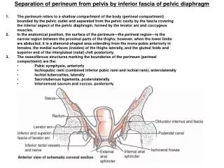
It is a muscular partition which separate the pelvic cavity from the anatomical perineum. • It consists of 3 sets of muscle on either side – pubococcygeus, iliococcygeus and ischiococcygeus, these are collectively called levatorani. • Upper surface is concave and slopes downwards, backwards and medially and is covered by parietal layer of pelvic fascia.
Inferior surface is convex and is covered by anal fascia. • The muscle with covering fascia is called the pelvic diaphragm. • Two gaps in the midline • hiatus urogenitalis • Hiatus rectalis
Structures inrelation to pelvic floor : Superiorly : • Pelvic organs : • Bladder, vagina and rectum • Pelvic cellular tissues - • pelvic peritoneum • Levatorani • Ureter and uterine artery Inferiorly : • Anatomical perineum
Nerve supply : Levator ani is supplied by 3rd/4th sacral nerve, inferior rectal nerve and perineal branch of pudendal nerve (S2, 3,4)
Functions : • To supports the pelvic organs – especially pubovaginalis • Protects the urogenital structures from increase in intra-abdominal pressure. • Facilitates internal rotation at the time of labour. • Helps anal sphincter in its action – puborectalis • It stabalises sacroiliac and sacroloccygeal joints – Ischiococcygeus • It stabalisesperineal body
Pelvic floor during pregnancy and parturation : Levator ani muscle hypertrophy and become more distensible and relax and favour the labour process.
The urinary bladder Hollow muscular organ Capacity – 450ml but can distend as much as 3-4 ltrs. Parts - Apex, superior surface, base, 2 inferiolateral surface, neck
Relations : Superior surface – peritoneum of the utero vesical pouch Base - supravaginal cervix and anterior fornix Inferior lateral surface - space of retzium.
It is divided into 2 areas (physiologic significance): 1)The base of the bladder- consists of the urinary trigone posteriorly and a thickened area of detrusor anteriorly. the three corners of trigone are formed by two ureteral orifices and the opening of the urethra into the bladder. the base receives α-adrenergic sympathetic innervation and is the area responsible for maintaining continence. 2)The dome of the bladder- remaining bladder area above the bladder base .it has parasymphathetic innervation and is responsible for micturition.
Structures : Outside to inside Outer visceral layer of the pelvic fascia. Muscle layer composed of muscle running in various directions. Near internal urethral opening the muscle fibres provide involuntary sphincter. Mucous coat is transitional epithelium with no gland. There is no sub mucous coat.
Blood supply : Superior and inferior vesicle arteries. The veins drain in to vesicle and vaginal plexus and to internal iliac veins. Lymphatics : Drain in to external and internal iliac lymphnodes.
Nerve supply : Sympathetic – pelvic plexus Parasympathetic pelvic plexus from the nerve erigentes (S2,3,4) Produces contraction of detrusor muscle and relaxation of internal sphincter. Sympathetic takes painful stimuli of over distention
Development : It is developed from upper part of urogenital sinus.
Pelvic ureter The ureter enters the pelvis in front of the bifurcation of the common iliac artery over the sacroiliac joint. As it courses downwards in contact with the peritoneum it lies anterior to the internal iliac artery and behind the ovary and forms posterior boundary of ovarian fossa.
On reaching the ischial spine, it lies over the pelvic floor and as it courses forwards and medially on the base of the broad ligament, it is crossed by the uterine artery anteriorly. Soon it enters the ureteric tunnel and lies close to the supravaginal part of the cervix, about 1.5 cm lateral to it.
After traversing a short distance on the anterior fornix of the vagina it courses in to the wall of the bladder obliquely for about 2 cm by piercing the lateral angle before it opens in to the base of the trigone.
Sructures: From outside to inward - Fibres derived from pelvic fascia - Muscle coat ; 3 layers - Mucous layer – transitional • Blood supply : segmental supply from – uterine,vaginal, vescical, middle rectal ( ant divisional of internal iliac) and superior gluteal arteries. Venous drainage by corresponding veins.
Lymphatics : Lower and middle part drain in to internal and external ilaiac and upper part drain in to lumbar lymph nodes. Nerve supply :Sympathetic from hypogastric and pelvic plexus and parasympathetic from sacral plexus. Development : It is developed as ureteric bud from mesonephric duct.