Download
1 / 42
430 likes | 982 Vues
Pelvic Floor Anatomy and Female Lower Urinary Tract Dysfunction. DR SHARON RASQUINHA. Genital support. Pelvic floor- Pelvic visceral attachment to pelvic walls through endopelvic fascia Levator ani muscles- a pelvic diaphragm with a cleft in anterior portion

E N D
Pelvic Floor Anatomy and Female Lower Urinary Tract Dysfunction DR SHARON RASQUINHA
Genital support • Pelvic floor- Pelvic visceral attachment to pelvic walls through endopelvic fascia • Levatorani muscles- a pelvic diaphragm with a cleft in anterior portion • Urogenital diaphragm connects perineal body to ischiopubicrami • Bulocavernous, ischiocavernous, sup.transverseperineal, anal sphincter m.
Levatorani muscle • Pelvic diaphragm composed of levatorani, coccygeus, obturatorinternus, and piriformis muscles • Levatorani consists of medial pubococcygeus and lateral iliococcygeus muscles • Pelvic diaphragm composed of levatorani, coccygeus, obturator
Functions of pelvic floor • Support pelvic and abdominal organs during stress of increased abdominal pressure • Allow for opening of the pelvic floor to accommodate excretory functions and parturition • Endopelvic fascia and visceral ligaments contains smooth muscles
Pelvic floor attachments • Pelvic floor support depends on its connection to the pelvic bones • An evolutionary solution for support of visceral organs • Pelvic floor muscles oppose gravity and increased abdominal pressures
Prevention of prolapse • Constriction –levatorani muscles constrict lumen of vagina • Suspension –cardinal ligaments & uterosacral ligaments, pubocervical fascia act to suspend cervix and vagina • Flap valve mechanism- anterior traction of levatorani m. and suspension of vagina in posterior pelvis
Attachments of pelvic floor • Constriction –levatorani muscles constrict lumen of vagina • Suspension –cardinal ligaments & uterosacral ligaments, pubocervical fascia act to suspend cervix and vagina • Flap valve mechanism- anterior traction of levatorani m. and suspension of vagina in posterior pelvis
Pelvic floor dysfunction • A variety of fascial and anatomic defects • Cystocele, rectocele, uterine prolapse, enterocele, vault prolapse • Adequate diagnosis and staging of pelvic floor dysfunction is essential
Diagnosis of pelvic floor dysfunction • Detailed physical examination • Pelvic ultrasound • Fluoroscopy of rectum & bladder • Magnetic resonance imaging (MRI)
Cystoele • Most Gr 1 and 2 cystoceles are asymptomatic • High grade cystoceles are associated with vaginal buldging, vaginal pressure, dyspareunia, UTI, obstructive voiding, urinary retention • A high grade cystocele may mask urethral hypermobility and stress incontinence
Enterocele • Simple enterocele • Complex enterocele- associated with vault prolapse and anterior or posterior vaginal prolapse • Cause vaginal pressure, dyspareunia, low back pain, constipation, symptoms of bowel obstruction
Rectocele • Defect of prerectal and pararectalfascia,andrectovaginal septum • Present in 80% asymptomatic patients • Vaginal mass,vaginal pressure, dyspareunia,constipation
Uterine prolapse • Laxity of uterosacral ligaments • May present with vaginal mass, dyspareunia, urinary retention, back pain • Grade 4 prolapse is associated with ureteral obstruction
Staging of pelvic organ prolapse • Stage 0 - no prolapse • Stage I - the most distal portion is >1cm above level of hymen • Stage II - The most distal portion is <1cm proximal or distal to plane of hymen • Stage III - The most distal portion is >1cm below plane of hymen • Stage IV - complete eversion of total length of lower genital tract
Ureter • A 25 – 30 cm long muscular tube transporting urine from kidney to urinary bladder. • Begins as a continuation of renal pelvis • It descends anterior to psoas major muscle (opposite the tips of lumbar transverse processes). • It crosses the end (bifurcation) of common iliac artery to enter the pelvis.
Runs downward in front to internal iliac artery, reaches ischial spine • Turns forward and medially , enters the upper lateral angle of urinary bladder • Near its termination, is crossed by the vas deferens • Passes obliquely through the wall of bladder for about ¾ inch before opening into the bladder cavity.
Bladder muscle contraction mechanically closes off ureteral orifice which prevents a reverse flow of urine toward the kidney
The ureter has constrictions at three points (sites of obstruction and stone impaction) • At the ureteropelvic junction • At the crossing of external/common iliac artery • At site of entrance to bladder
Blood supply • Ureter is supplied by multiple arteries throughout its course • From above downward, these are:: • Renal artery • Gonadal artery • Common iliac artery • Internal iliac arter
Urinary bladder • Located immediately behind the pubic symphysis • Shape and relations vary according to the amount of urine it contains • An empty bladder: • In adults, is entirely a pelvic organ; as it fills, rises up into the hypogastric region. • In young children, it projects above the pelvic inlet
Apex • Directed forward • Lies behind the upper margin of the symphysis pubis • Is connected to umbilicus by the median umbilical ligament (remnant of urachus)
Base or Posterior surface • Triangular in shape • Upper part covered by peritoneum • Lower part related to: • In males: vas deferentia and seminal vesicles • In females: vagina Infrolateral surfaces: • Related in front to the retropubic pad of fat & the pubic bones • Posteriorly lie in contact with the obturatorinternus above and levatorani below
Neck: • Lies inferiorly, and is the most fixed part of the bladder • Is related to lower border of symphysis pubis • The circular muscle fibers thickened to form the sphincter vesicae
Interior of urinary bladder • Mucous membrane thrown into folds except in the triangular region in the base of bladder, between the openings of the two ureters and the urethra. This region is called the ‘trigone’.Here The mucous membrane is always smooth even when the bladder is empty
Blood supply: • Arterial supply: from internal iliac artery • Venous drainage: into internal iliac vein • Lymphatics:into internal iliac lymph nodes
The nerves form the vesical nerve plexus that contains: • Sympathetic fibers derived mainly fromL1,2 • Parasympathetic fibers derived from pelvic splanchnic nerves S2,3,4 • Sensory fibers from the bladder are visceral and transmit pain sensation resulting from overdistention
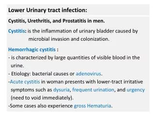
Urethra • Extends from neck of urinary bladder to open externally through the external urethral orifice (anterior to the vaginal opening) • Has only urinary function