Physiotherapy of Lower Urinary Tract Dysfunction
640 likes | 1.06k Vues
Physiotherapy of Lower Urinary Tract Dysfunction. Hann-Chorng Kuo Department of Urology Buddhist Tzu Chi General Hospital. Lower Urinary Tract Dysfunction. Urinary Incontinence Stress, urge, or mixed incontinence Frequency urgency syndrome Spastic urethral sphincter syndrome

Physiotherapy of Lower Urinary Tract Dysfunction
E N D
Presentation Transcript
Physiotherapy of Lower Urinary Tract Dysfunction Hann-Chorng Kuo Department of Urology Buddhist Tzu Chi General Hospital
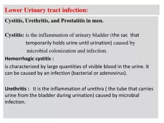
Lower Urinary Tract Dysfunction • Urinary Incontinence Stress, urge, or mixed incontinence • Frequency urgency syndrome • Spastic urethral sphincter syndrome • Poor relaxation of urethral sphincter • Pelvic pain syndrome • Chronic eliminative syndrome
Therapeutic modalities • Medical treatment • Surgical treatment • Behavioral therapy • Physiotherapy Electrical stimulation Biofeedback PFMT Neuromodulation Neurostimulation
Functional Electrical Stimulation • Restoration of normal physiological reflex mechanisms in abnormal nerves and muscles • Black torpedo fish in 46 AD • Bors (1952) electrostimulation of pelvic floor • Caldwell (1965) anal and urinary incontinence by electrical stimulator • Alexander & Rowan (1968) electrodes on vaginal pessary • Suhel (1975) integrated automatic vaginal stimulator
Neuromuscular Electrical stimulation • Excitation of peripheral nerves using short pulses, adequate intensity and duration • Current amplitude (intensity) • Pulse width (duration) • Pulse rise time • Pulse repetition rate (frequency)
Muscle Fatigue • Skeletal muscle is composed of aerobic slow contracting motor units and anaerobic fast contracting units • Resistance to fatigue is inversely correlated to aerobic oxidative capacity • At high frequency electrical stimulation the muscle fatigues rapidly due to impaired neuromuscular transmission and sarcolemmal excitation
Skeletal muscles • Motor striated muscles are composed of slow, intermediate, and fast contracting muscles, fast muscle has 10-20 times more contraction force than slow fibers • Intramural urethral sphincter – small slow muscle fibers • Periurethral pelvic floor muscles – all types of muscles • Provocative situation – fast fibers of PFM action to close urethra
Muscle Activity • Plasticity of metabolic and functional properties of muscles • Following denervation, muscles lose enzymatic difference • Immobilization induced muscle atrophy • Disuse atrophy the muscle response is weak and rapid fatigue
Chronic nerve stimulation • To modify physiologic and metabolic characteristics of normal & atrophied muscles • Transform fast to slow myosin subunits that are more fatigue resistance • Anaerobic fast muscle turns into slow muscle with a high capacity for energy supply by aerobic oxidative process • Increase myoglobin and mitochondria content • Increase in capillary density
Muscle Transformation after Nerve Stimulation • Transformation of fast to slow twitch muscles is progressive with the duration of stimulation • The most extensive changes occur between 60 and 90 days • The total number of fibers remains constant • Intermittent phasic high frequency stimulation (40 to 60 Hz) induces transformation similar to that after low-frequency (10Hz) stimulation • The reverse process occurs by inactivity and chronic immobilization
Pelvic Floor Muscle Stimulation • Induces a reflex contraction of striated para- and periurethral muscles and a simultaneous reflex inhibition of detrusor contraction • A sacral reflex arc and peripheral innervation must be intact • No effect can be expected in complete lower motor neuron lesions
Nerve Stimulation for Urethral Closure • Direct stimulation of efferent pudendal nerves • Activation of efferent hypogastric fibers can contract smooth urethral muscles • Efferent stimulation of pelvic nerves can increase intraluminal urethral pressure and increase urethral length • Stimulation of pelvic floor afferents from anogenital muscles and mucosa may activate pelvic floor muscles through reflex connection
Nerve Stimulation for Bladder Inhibition • A feedback system is present in micturition process • Detrusor instability may be caused by ineffective inhibition by sphincter • Intravaginal or pudendal nerve stimulation of sufficient intensity causes a complete bladder relaxation • The higher intensity the more efficient bladder is inhibited via spinal reflex mechanism
Nerve Stimulation for Bladder Relaxation • Maximal bladder inhibition is obtained at 2x to 3x of threshold intensity • Relaxation of detrusor is accompanied by tightening of bladder neck fibers • Detrusor inhibition after nerve stimulation may be caused by balance between cholinergic (M2,3-receptors) and beta-adrenergic (B3-receptors) neurotransmission • After maximal stimulation, high beta-adrenergic activity and decreased cholinergic activity in rabbit detrusor strips
Chronic Pelvic Floor Stimulation • Chronic long-term stimulation of anal and urethral sphincters applies relatively weak electrical impulses for 3 to 12 months • Fast motor units are recruited first • Increase frequency of slow-twitch fibers • Accelerated sprouting of surviving motor units of partially denervated pelvic floor muscles • High frequency (25-50 Hz) is advised in treating stress incontinence
Selection of Electrical Parameters • Patient adapt to current intensity within a few minutes • The stimulation is constructed to increase current intensity from 0 to maximum within a few minutes • A pulse length of 0.5 to 1.0 minutes is optimal to muscle contraction • Biphasic pulses give 30% to 40% better therapeutic response than monophasic pulses
Selection of Frequency of Electrical Stimulation • Maximal detrusor inhibition is obtained with a frequency of 5 Hz • No difference in MUCP change in the range of 10- 50 Hz • Good therapeutic results in stress and urge incontinence with a fixed frequency of 25 Hz • Intermittent ES is superior to continuous ES to avoid muscle fatigue during long-term stimulation • The most effective rest period is 3 times longer than active period
Functional ES for Stress urinary incontinence • Successful pelvic floor stimulation was reported in 50- 92 % women with incontinence • Patients without previous incontinence surgery have the best result • Urodynamic parameters change little after functional ES for SUI • Patients with SUI may have a better pelvic floor muscle contractility after ES that results in increased urethral resistance during stress
Long-term electrostimulation • At least 6 to 8 hours daily ES is needed either anally or vaginally • A treatment period of 3 to 6 months is necessary to achieve success • Kegel exercises should be followed after discontinuing FES to keep pelvic floor muscles in optimal condition • Treatment combined with estrogen is recommended in menopause women • Mechanical vaginal mucosal irritation may occur in atrophic vaginitis
Short-term Maximal stimulation • Intact reflex arc must be present • Maximal ES can inhibit overactive detrusor muscle, can be an alternative in treating detrusor overactivity and urge incontinence • 5 to10 Hz can give optimal inhibitory effect • The current intensity is successively increased below pain level of patient • Duration of maximal ES is 15 to25 minutes, 4 to 10 repetitions daily for 2 to 3 days
Therapeutic Results after Short-term electrostimulation • Successful maximal ES for pelvic floor in female urge incontinence was reported to be 52 to 92% • A recurrence rate of 25% after discontinuing maximal ES in urge UI • Recurrence rate of 15% within 1 year • Success rate of 75% in recurrent urge urinary incontinence • Repeat stimulation is needed for recurrence
Electrical Stimulation for SUI • Transvaginal ES has been used for genuine SUI, urge and mixed urinary incontinence • Reported efficacy ranges 35 to70% • A placebo-controlled study revealed after 15-week treatment course, pad usage diminished by >50% in 62% women compared to 19% in sham device, incontinence episode reduced >50% in 48% women compared to 13% in sham device
Transvaginal electrical stimulation • Low frequency (20 Hz) was applied • Contrasting data of effects on genuine SUI • Transvaginal ES is effective in urge UI • First line treatment for women with pure urge incontinence • For the women with mixed type UI who does not wish to undergo PME or surgery
Transvaginal electrical stimulation for Urge incontinence • Leach reported 6% after long period of stimulation • McGuire observed improvement in 93% women with urge incontinence • Plevnik found 52% improved (30% cured) in pure urge incontinence • Brubaker used 20 Hz frequency current and cured 49% with urodynamic DI • Smith found ES reduced urine loss by 50% in 20women • Sand reported 38% success rate in 20 women with DI
Contraindication of ES • Heart pacemakers • Pregnancy women • Urethral obstruction and overflow incontinence • Complete peripheral denervation • Urinary tract infection • Uterine prolapse or high grade cystocele • Low compliance and cooperation of patient
Biofeedback • Detectable or measurable response: bladder pressure or pelvic floor muscle activity • A detectable response • A perceptible cue : sensation of urge or tightness • Active involvement of a motivated patient
Biofeedback for LUTD • Fail to inhibit detrusor contraction • Fail to adequately contract striated urethral sphincter of the pelvic floor • Failed to relax the urethral sphincter or pelvic floor muscles during micturition • Chronic pelvic pain due to hypertonicity of pelvic floor muscles
Cystometry biofeedback for urge incontinence • For women who failed electrical stimulation, were intolerant to anticholinergics, • Urodynamic detrusor overactivity was proven • Performed several voluntary PFMC at episodes of DI while watching CMG tracing and EMG activity • Try to inhibit urge incontinence as longer duration as possible at home
Bladder biofeedback • Train patients to inhibit detrusor contraction voluntarily and to contract periurethral muscles selectively • Bladder pressure biofeedback to treat urge incontinence by watching intravesical pressure rise during CMG • 81% improvement rate was reported and 36% success rate at 5 year follow-up
Pelvic Floor Muscle Biofeedback • Vaginal manometry – by perineometry Kegel reported a 90% improvement rate • Vaginal electromyography – in 8 week program 80% younger and 67% older group reported no more incontinence • Anal sphincter biofeedback – by perineal surface EMG or rectal probe
Pelvic floor hypertonicity & overactivity Etiology • Persistence of a reaction phase to noxious stimulus of LUTS (e.g. inflammation, infection, irritation, post-surgery) • learned dysfunctional voiding behavior • Persistent transitional phase in the development of micturition control • Sexual abuse
Clinical presentation • Dysfunctional voiding Increased pelvic floor activity during voiding Urgency frequency, poor stream, intermittency, hesitancy • Urinary retention • Constipation • Pelvic or perianal pain Certain pelvic pain (e.g. interstitial cystitis, prostatodynia, urethral syndrome) is associated with pelvic floor hypertonicity
-incontinence -reflux -mucosal ischaemia -diet regulation -drinking and voiding chart -pharmacotherapy Bladder dysfunction Overtraining of the pelvic floor muscles Pelvic floor dysfunction Biofeedback electrical stimulation manual technique -milk-back of urine -residual urine -pelvic pain
Aims of physical therapy • To improve dietary and micturition routine • To improve proprioception and body awareness of PF: focus on relaxing the PF and voluntary sphincter control • To decrease any associated hypertonicity or pain in the PF • To optimize functional use of PF
Evaluation A complete history • Frequency /volume chart for 3 days Neurological examination (lower quarter) • proprioception, sensation • Peripheral reflexes Physical examination PF function: Rectal /vaginal tone, contractility, endurance, ability to contract and relax PF voluntarily, relation between PF & adjacent pelvic viscera • pelvic pain: trigger point, tenderness • Sacroiliac & coccygeal position /mobility
Behavioral modification • Instruction on urinary system and PF dysfunction • Diet: avoid bladder stimulants, high fiber adequate daily intake of water • General recommendations for changing wrong voiding behavior take time for micturition, do not push Instruct a proper toilet posture: sit for voiding every time (men also) no straining timed voiding (3 ½~4 hours)
Manual technique To restore sacroiliac & sacrococcygeal alignment To improve proprioceptive awareness • Muscle energy technique • Proprioceptive technique: direct pressure, tapping, use of stretch reflex To decrease tension and promote relaxation of the musculature • Massage • Trigger point pressure • Myofascial release
Clinical effectiveness • Standford CA internal myofascial release, 18 sessions hypertonus & pain in type III chronic prostatitis • Jerome MW myofascial release, 8-12 weeks 83% urgency-frequency syndrome symptom relief & hypertonus 70% interstitial cystitis
Pelvic floor exercise (PME) with EMG biofeedback • Convert pelvic floor/urethral sphincter activity into visual or auditory signal • Goal: • to help identify pelvic floor musculature • to perceive difference between contraction, relaxation, and straining • to voluntary relax & control pelvic floor
EMG biofeedback: children with dysfunctional voiding • Anal plug or surface electrode on perineal skin • Protocol: a short submaximal contraction (3 sec) a prolonged relaxation (30 sec) for 30 times with diaphragmatic breathing progress: increase holding time (10 s) followed by prolonged relaxation (30 s)
PME with EMG biofeedback • Intravaginal/ intra-anal EMG sensor • Glazer Protocol • One minute rest, pre baseline • Five rapid contraction (flicks) with 10-s rest between each • Five 10-s contractions with 10-s rest between each (tonic) • A single endurance contraction of 60-s • One minute rest, post baseline