Download
1 / 22
220 likes | 429 Vues
Muscle Physiology. Dr Taha Sadig Ahmed. RMP = -90 mV ( same as in nerves ) Duration of AP = 1-5 ms ( longer duration than nerve AP , which is usually about 1 ms ) . Conduction velocity (CV) in a muscle fiber ( cell) = 3-5 m/s ( slower than big nerves ). The Muscle Action Potential.

E N D
Muscle Physiology Dr TahaSadig Ahmed
RMP = -90 mV ( same as in nerves ) Duration of AP = 1-5 ms ( longer duration than nerve AP , which is usually about 1 ms ) . Conduction velocity (CV) in a muscle fiber ( cell) = 3-5 m/s ( slower than big nerves ) . The Muscle Action Potential Amplitude إرتفاعه RMP= - 90 mV
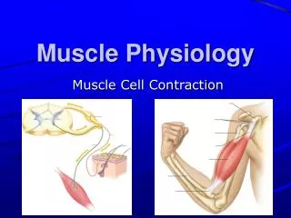
Muscle Contraction There are 4 important muscle proteins : A/ two contractile proteins that slide upon each other during contraction : • Actin • Myosin B/ And two regulatory proteins : • Troponin excitatory to contraction • Tropomyosin inhibitory to contraction
The EPP at the motor end-plate triggers a muscle AP The muscle AP spreads down inside the muscle through the Transverse Tubules ( T-tubules ) to reach the Sarcoplasmic Reticulum (SR) . In the SR the muscle AP opens calcium channels ( in the walls of the SR) calcium passively flows out ( by concentration gradient ) of the SR into muscle cytoplasm Ca++ combines with Troponin Tropomyosin gets moved Myosin heads combines with Actin
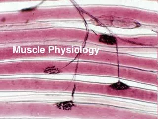
Skeletal muscle is made up of many cylinderical ,multinucleated muscle cells ( fibers) • The fibers ( cell ) can be 10 to 100 ten micron in diameter , and can be hundreds of centimeters long. • & is covered by a cell-membrane called Sarcolemma. One muscle cell ( fiber ) Sarcolemma
Each cell contains between few hundreds to a few thousands Myofibrils The myofibril is striated cand has dark bands called A-bands) and light bands called I-bands (I-bands). Each Myofibril Is made of 3000 Actin filaments And 1500 Myosin filaments .
Each myofibril is striated • It is made of Sarcomeres • And each sarcomere is limited by two Z-lines (Z disk) • The sarcomere contains both the (i) A band and (ii) I band light Z-lines
A-bands consist mainly of thick filament Myosin • The ends of Actin are Z-Discs(Z-lines ). • I-bands consist of thin filament Actin. • The part of the Myofibril lying between two Z-discs is called Sarcomere .
Sliding Filament Mechanism When contraction takes place Actin & Myosin slide upon each other , & the distance between two z-discs decreases This is called Sliding Filament Mechanism . Z-line come closer together I-band gets smaller , and eventually may disappear A-band does not become smaller or bigger
Actin is made of globularprotein callledG-actinG-actins are attached together to form F-actin strand ( chain ) Each two strands wind togetherto form double helix called Actin Filament Tropomyosinlies in the groove between the F-actin strands to cover the active sites on actin that bind the head of myosinTroponinis attached to tropomyosin and to actin Two Actin filaments Groove between the 2 F-actin strands Tropomyosin covering active sites on Actin
Attachment of Ca++ to Troponin initiates tcontractionand when is activated by Ca++ it will move the Tropomyosin away from the active sites on actinn & expose them for Myosin .> then myosin head will immediately attach to these actin active sites > when the myosin head attaches to actin it forms a “ cross-bridge”
Myosin • Each 200 myosin molecules aggregate to form a myosin filament , from the sides of which project myosin heads in all directions .
Myosin • Each Myosin molecule has • (1) Head • ( 2 ) Tail • (3) Hinge (joint ) • Furthermore , each myosin head contains • (1) ATP-binding site , & • (2) ATP-ase enzyme .
Attachment of Myosin to Actin activates the enzyme ATPase in the Myosin Head • ATPasebreaks down ATP releasing energy • This energy is used in the “Power Stroke ” to move the myosin head leading to pulling & dragging of actin • sliding of actin on myosin • The “ power stroke ” means tilting of the Myosin cross-bridge and dragging ( pulling ) of actin filament
Then , on order to release the head of Myosin from Actin , a new ATP is needed to come and combine with the head of Myosin . A new ATP binding to Myosin head is essential for detachment of Myosin from Actin
Q: What is Rigor Mortis ? • Q: ATP is neede for 3 things : what are they ? • Q: Is muscle relaxation a passive or active process ? Why ? • Q: What happens to A-band and I-band during contraction ? • Q: Ca++ is needed in nerve & muscle : when and where ?
Why do we need the ATP in contraction ? • ATP is needed for 3 things : • (1) Power stroke. • (2) Detachment of myosin from actin active sites . • (3) Pumping C++ back into the Sarcoplasmic reticulum