Download

1 / 33
330 likes | 416 Vues
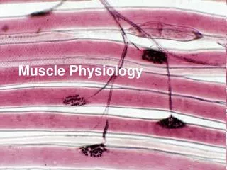
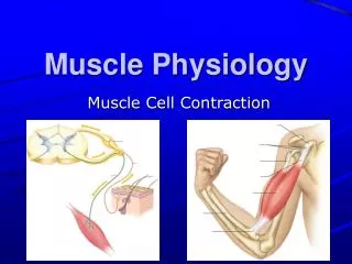
Muscle Physiology. Connective Tissue Components. Muscle cell = muscle fiber Endomysium – covers muscle fiber Perimysium – binds groups of muscle fibers ( fasicles ) Epimysium – covers the entire muscle Tendon – fibrous tissue that connects muscle to bone

E N D
Connective Tissue Components • Muscle cell = muscle fiber • Endomysium – covers muscle fiber • Perimysium – binds groups of muscle fibers (fasicles) • Epimysium – covers the entire muscle • Tendon – fibrous tissue that connects muscle to bone • Aponeurosis – broad, flat sheet of connective tissue • Fascia – fibrous CT surrounding muscle and tendon
General Function of Muscles • Movement • Excitability • Contractility • extensibility • Heat Production • Posture
Overview of Muscle Cell • Muscle cell = muscle fiber • Sarcolemma = plasma membrane • Sarcoplasm = cytoplasm • Sarcoplasmic reticulum (SR) = network of tubules and sacs • Multi-nucleated, multiple mitochondrion • Bundles of myofibrils extend lengthwise & fill sarcoplasm • Composed of thick and thin myofilaments
Sarcomere • Contractile unit of a muscle fiber • each myofibril consists of many sarcomeres • Z line • Anchors thin filaments • Boundary of sarcomere • M line – anchors thick filaments • A band: segment of thick & thin filaments • I band: segment of thin filaments • H zone: where thin and thick filaments will not overlap (only thick)
Sarcomere cont… • Elastic filaments – connect thick filaments to Z line • T (transverse) tubules – allows impulses traveling along sarcolemma to move deeper within the cell • Triad – t tubule sandwiched between sacs of the SR • Allows impulses traveling along a t tubule to stimulate sacs of the SR
Myofilaments • Myofibrils – made up of 1000s of thin and thick myofilaments • Thin filaments • Actin • Tropomyosin • Troponin • Thick filaments • myosin
Muscle Excitation • Nerve impulse reaches the end of a motor neuron releases acetylcholine (Ach) • Ach diffuses across the neuromuscular junction and binds with the receptors on the motor endplate
Muscle Contraction • Impulses travel along the sarcolemma t tubules sacs of SR • Ca2+ is released into the sarcoplasm binds with troponin on thin myofilaments • Tropomyosin shift to expose actin’s active site • Energized myosin heads bind with myosin’s active site and pulls thin filament towards center of sarcomere • Requires ATP
Muscle Relaxation • Nerve impulse is complete Ca2+ is pumped back into the sacs of the SR • Ca2+ is stripped from the troponin tropomyosin covers the actin’s active site • Myosin heads can no longer bind with myosin muscle fiber returns to its resting length
Rigor Mortis • “stiffness of death” • SR releases excess Ca2+ myosin heads bind with actin’s active sites contraction of myofilaments • Lack of ATP after death causes cross bridges to “stick”
http://highered.mcgraw-hill.com/sites/0072507470/student_view0/chapter9/http://highered.mcgraw-hill.com/sites/0072507470/student_view0/chapter9/
Sliding Filament Theory In fully contracted muscle: • H zone disappears • I band narrows • A band remains the same
Energy for Contractions • Hydrolysis (breakdown) of ATP • ATP ADP (breaking high energy bond btwn 2nd and 3rd phosphate groups) • ATP binds myosin head moves to resting position (11-7A) • Breakdown of ATP allow myosin head to bind with actin and perform “power stroke” (11-7B-D) • ATP binds to return myosin head back to resting position
Alternate Source of Energy • ATP must be continually re-synthesized • Breakdown of creatine-phosphate (CP) provides energy for ATP re-synthesis • Catabolism of food provides energy for ATP and CP synthesis
Oxygen & Glucose • O2 and glucose are the starting materials for cellular respiration (process that makes ATP) • During rest oxygen is stored in myoglobin • Supplies muscle fibers with oxygen during period of exercise • High amounts of myoglobin = red fibers = slow twitch fibers • Low levels of myoglobin = white fibers = fast twitch fibers
Aerobic vs. Anaerobic Respiration • Aerobic Respiration • Oxygen-requiring process • Produces maximum amount of ATP from one glucose molecule • Anaerobic Respiration • Does not require oxygen • Short-term, rapid process to re-synthesize ATP • Produces lactic acid • Burning/soreness in muscles
Heat Production • Some energy from catabolic processes is lost as heat • Muscle release massive amts of heat • Thermoreceptors sense decrease in body temp hypothalamus integrates information signal sent to skeletal muscle to contract shivering homeostatic balance is maintained
Motor Unit • Motor unit = motor neuron + muscle fibers it attaches to • Motor neurons can innervate few to 100s of muscle fibers • A lower number of muscle fibers within a motor unit = more precise movement • Ex: hand vs abdomen
http://natchem.files.wordpress.com/2009/11/motor-unit-lg.jpg
Isotonic vs Isometric Contractions • Isotonic – tension remains the same; length of the muscle changes • Concentric contraction: muscle shortens (contracts) • Eccentric contraction : muscle lengthens • Isometric – tension changes; length of the muscle remains the same • Myosin heads unable to move thin filaments • Static tension
Smooth Muscle Contractions • Small tapered cell w/ single nuclei • No t-tubules; loosely organized SR • No sarcomeres • Contract to shorter lengths • Myofilaments crisscross (balled up appearance when contracted) • Calcium binds to calmodulin
http://www.cytochemistry.net/microanatomy/muscle/smooth1.jpg
Smooth Muscle Tissue Types 1. Visceral • Gap junctions connect smooth muscle fibers into sheets • Forms inner muscular layer of hollow structures • Exhibits autorhythmicity • Peristalsis, excretion of urine, childbirth, mixing of stomach contents 2. Multiunit • Composed of many single-cell units • Ex: arrector pili muscles, lines blood vessels