Understanding Muscle Physiology: Structure, Function, and Fibers
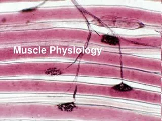
Muscles are composed of fibers, which are individual cells. Fascia connects these fibers, forming muscles and separating them from one another. There are three layers of connective tissue: epimysium (surrounds the entire muscle), perimysium (surrounds and separates fascicles), and endomysium (surrounds each muscle fiber). The sarcolemma is the membrane of a muscle fiber, while the cytoplasm is called sarcoplasm. Myofibrils within the fiber consist of actin (thin filaments) and myosin (thick filaments), arranged into sarcomeres, the basic functional units responsible for muscle contractions.


Understanding Muscle Physiology: Structure, Function, and Fibers
E N D
Presentation Transcript
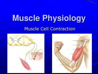
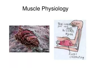
Muscle Physiology How They Work
Muscle Fibers • Muscles are made of many individual cells called fibers • The Fascia connects the individual fibers to form a muscle and it separates muscles from each other.
Muscles have 3 layers of C.T. • Epimysium: outermost layer that surrounds the whole muscle • Perimysium: Surrounds and separates the fascicles • Endomysium: surrounds each individual muscle fiber
Muscle fiber membrane: • The plasma membrane of a muscle fiber is called the sarcolemma • The cytoplasm is called the sarcoplasm
Inside the sarcolemma • The many parallel fibers inside the muscle cell are called myofibrils. • Each myofibril is made of protein filaments called myofilaments. • The thin filaments are actin. • The thick filaments are called myosin.
What is a sarcomere? • Actin and Myosin filaments are arranged in an overlapping pattern of light ("I" bands) and dark ("A" bands). • In the middle of each "I" band is a line called a "Z" line. • The section of a myofibril from one Z-line to the next Z-line is the SARCOMERE.
The arrangement of the sarcomeres produces the familiar striations of skeletal muscle.