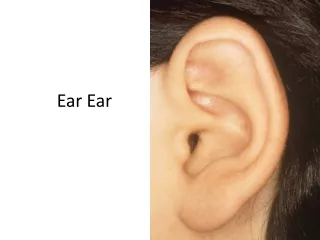
Common Ear Conditions
Common Ear Conditions. Medway VTS 2013. Plan. Consider a few common presentations in general practice related to ears . Examining the ears . Ear Wax and syringing . Otitis Externa . Otitis Media( Acute and Chronic) . Eustacian tube dysfunction . Perforations ( Safe vs. Unsafe)
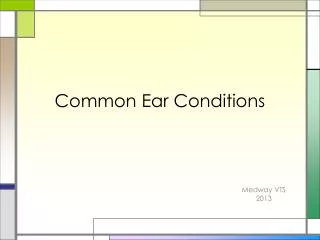

Common Ear Conditions
E N D
Presentation Transcript
Common Ear Conditions Medway VTS 2013
Plan Consider a few common presentations in general practice related to ears . Examining the ears . Ear Wax and syringing . Otitis Externa . Otitis Media( Acute and Chronic) . Eustacian tube dysfunction . Perforations ( Safe vs. Unsafe) Treatment Few questions
ENT Examination You tube video of ENT examination in an OSCE situation.http://www.youtube.com/watch?v=mDbwAPr5RvU Ear examination- You tube video http://uk.youtube.com/watch?v=I3sa2W83iuo&NR=1 NB: The canal may be partly straightened by pulling the pinna backwards and upwards during examination In infants pull the pinna more horizontally backwards as the shape of the ear canal is different
Normal Attic Consider the malleus as an arrow; pointing in the forward direction The normal tympanic membrane should appear: . pearly grey . have a light reflex . generally concave . With a visible malleus Anterior direction Anterior Posterior Inferior
Ear Drum-normal Landmarks An annulus fibrosus or more commonly referred to as the eardrum margin. This is important. Note how smooth and how ever so slightly blurry it is. Umumbo - the end of the malleus handle and usually marks the centre of the drum Lr light reflex or Cone of light –is usually seen antero-inferioirly At Attic also known as pars flaccida. Any perforations here are serious and need referral.
Examine out to in External: Pinna (shape, colour, position, tenderness, haematoma) Mastoid Internal: The Canal ( skin, spores, foreign bodies, discharge, debris, wax) The Tympanic membrane (look ant, post, superior/ attic and inferior of malleus) . Colour( opaque, white, red, patches & translucency) . Retraction( landmarks behind it more visible) . Perforation ( safe/ unsafe) . Discharge (mucopurulent) Behind the Eardrum . Fluid behind the drum( meniscus, colour, bubbles) . Any red bits( glomus tumour, granulations or blood?, white- cholesteotoma)
Ear Wax Wax is produced in the outer half of the ear canal and migrates outwards along with the canal skin. Inappropriate instrumentation can cause impaction. Wax impaction can cause hearing loss, pain, tinnitus, vertigo, or chronic cough but not usually discharge. Sudden expansion after getting water in can cause sudden deafness or pain, but needs careful exclusion of other pathology behind it e.g. infection Crayon in a child’s ear
Management: Cotton buds are not your friend If Symptomatic – topical meds Different preparations available none superior to other. Sodium bicarbonate drops might be better at disintegrating wax, but can cause dryness of the canal and/ or irritation . Instructions for use: e.g. Olive oil? . Syringing . When to refer to ENT clinic: . Patients known to have a tympanic membrane perforation or previous ear surgery (need microsuction), only hearing ear . Syringing fails . Causes pain or vertigo, . Hearing loss persists after wax removal.
Otitis Externa Infection of the external auditory canal. Mediterranean ear/Swimmers ear Usually unilateral Gradual onset pruritis, pain, hearing loss, and ear discharge which varies in consistency and colour. The pt is usually well. Can result in a featureless ext aud canal Risk factors: trauma, water, Immunosuppression, eczema Can be fungal- spores might not always be visible If treatment fails or otitis externa recurs frequently consider sending an ear swab for bacterial and fungal microscopy and culture
Management Remove or treat any precipitating or aggravating factors A topical ear preparation for 7 days. Options include preparations containing: a. Both a non-aminoglycoside antibiotic + a corticosteroid e.g. flumetasone–clioquinol (Locorten–Vioform®) ear drops. b. Both an aminoglycoside antibiotic and a corticosteroid (contraindicated if the tympanic membrane is perforated). c. Topical preparations containing only an antibiotic (gentamicin ear drops are contraindicated if the tympanic membrane is perforated). d. Antifungal or ? something containing all three Aural toilet: if earwax or obstruct topical medication (may require referral). Provide appropriate self-care advice
Malignant Otitis Externa "Malignant" otitis externa is a severe infection due to Pseudomonas aeruginosa and anaerobes causing osteomyelitis of the skull base characterised by severe pain, involvement of the floor of the ear canal, sometimes with granulation tissue. If untreated, it can involve the cranial nerves and brain. Facial nerve palsy occurs in 50% of patients, IX to XII may also be involved. immunocompromised patients, especially elderly diabetics. It may be life threatening. What to look for:Elderly, DM, earotalgia, otorrhoea, hoarseness, puffiness , trismus, failure to respond to drops, granulations, CN palsies etc Mx: -Refer -Intensive local and systemic antibiotics against Pseudomonas are required if malignant otitis externa is present, e.g. ciprofloxacin or ceftazidime, plus suitable anaerobic cover e.g. metronidazole.
Question 1 23 yr old man, 4 days Hx of itchy sore Rt ear; returned recently from holiday in Spain O/E= Rt ext auditory canal is inflamed but no debris seen. T.membrane is visible and unremarkable. What is the most appropriate management? A. Topical corticosteroid + Aminoglycoside B. Topical corticosteroid C. Tell him serves him right for going on a holiday while you work! D. Topical corticosteroid +Clotrimazole E.. Oral Flucloxacillin
Answer 1 Correct Answer is A. Dx- Otitis externa- Topical antibiotic or combined Antibiotic + corticosteroid preparation
Question 2 53 year old man, fastidiously clean, previously normal hearing, currently recent onset ‘strange sensation in me ear!’ + slightly reduced hearing ‘have been trying to pop them’. The cone of light is normal, but what is this? • Normal ear drum • Otitis Externa secondary to ear buds use • Serous Otitis Media • Time waster/ Hidden agenda
Answer 2 Serous Otitis media because of Eustacian tube dysfunction Has normal cone of light, mild redness externally likely normal, fluid level, and mildly retracted ear drum
Question 3 A 28 year old woman presents with a 5 day Hx of pain in her Rt ear, reduced hearing, and yellow coloured discharge Q. What is the likely diagnosis a. Acute Otitis Media b. Acute Otitis Externa c. Chronic Suppurative Otitis media
Answer 3 Answer is Acute Otitis Externa
Question 4 Which of the following statements about otitis externa is correct? • You should avoid removing canal debris • Its common in people not wearing ear protection while working with loud power tools. • It may result in a featureless tympanic membrane d. It is usually due to a Staphylococcus aureus infection
Answer 4 Correct answer- It may result in a featureless tympanic membrane Commonest causative organism for infective otitis externa is Pseudomonas Could be difficult to eradicate in someone wearing ear protection in certain occupations e.g. forge/factory workers
Question 5 Which of the following statements about the use of topical eardrops is correct? a. Only use topical ear drops if the tympanic membrane is visible b. Topical eardrops are contraindicated in children under the age of 12years c. Topical eardrops cannot be used in the presence of a perforated tympanic membrane d. Topical eardrops can worsen otitis externa e. If its difficult putting them in your ears, they are equally effective putting them in your nose.
Answer 5 Correct answer- Topical eardrops can worsen otitis externa if there is sensitivity to them The use of ototoxic drops in the presence of a perforated tympanic membrane is controversial due to reports of sensorineural hearing loss as a result of their application. Reports of this association are rare and often the validity of such reports is questionable. Certainly the risks of sensorineural hearing loss or of major complications of otitis media are of more significance. Limiting the course of treatment and ensuring that they are not used in healthy ears can reduce any potential risks from the administration of ototoxic medicines.
Otitis Media Can be acute or chronic Can be with or without serous effusion (acute or chronic) Can be Acute or chronic suppurative Can co-exist with Otitis externa Otitis media with serous effusion= Glue Ear
Acute Otitis Media Common in children Unwell/pyrexia, otalgia/discharge there may be tenderness over the mastoid discharge in meatus loss of outline of drum and landmarks TM: red, bulging,oedematous or perforation. Mostly viral but can be Streptococcus/Haemophilus Risk factors: Passive smoker Male Family history of otitis media. In day care On formula feed
Current evidence for AOM 80% of children get better by day 3 without antibiotics ‘It is reasonable to prescribe analgesia.’- Antibiotics should not be used routinely and prescribing them just increases parental belief and re-attendance rates Use def scripts if necessary Adenoidectomy, as the first surgical treatment of children aged 10 to 24 months with recurrent acute otitis media, is not effective in preventing further episodes. Neither is Chemoprophylaxis. Current Evidence for CSOM Randomised controlled trials (RCTs) found limited evidence that topical quinolone antibiotics versus placebo improved otoscopic appearances. RCTs found no clear evidence of significant differences between topical antibiotics. No benefits from anything else.
AOM continued.. Analgesia: For most children helps most. Antibiotics should not be routinely prescribed for uncomplicated AOM. Some children may significantly benefit from antibiotics – ill. Choice of antibiotic: Amoxicillin is the usual first-line for 5 days. If severe symptoms present, or there has been a previous episode of AOM within the last month, use high doses Erythromycin or Clarithromycin are alternative antibiotics if allergic to penicillin
AOM contd 2…. A good compromise is to use issuing a delayed/deferred prescription to be redeemed within 72 hours only if the condition has not adequately improved. Active Follow up for: . under 2 years of age. . systemic symptoms such as high temps (> 39°C) or vomiting. . There is discharge from the ear. Visualisation of the tympanic membrane can be difficult. Re examine after 2 weeks to assess the integrity of the membrane and to check for complications. If there is a perforation still present, monitor the situation and consider referral if it has not healed after 6 weeks.
Serous Otitis Media/Secretory • Glue ear, commonest cause of deafness, and the commonest indication for surgery, in children. • The condition is most frequent in early childhood, • Peaks prevalence at 2 and 5 years. • Half of 3-year-olds have at least one effusion in a year, and in the UK, 1 in 200 children is operated on for the condition. • Ninety thousand operations are performed in England and Wales annually, at an estimated cost of £30 million
Hearing tests? A hearing test is not appropriate at the initial presentation if there is no evidence of significant hearing loss or developmental delay. If signs and symptoms of OME continue, hearing should be assessed after 3 months, where OME can be regarded as persistent.
Otitis media+effusion-Glue ear Features Dull retracted TM May show air-fluid level Conductive hearing loss Common in children; often after AOM and can persist for weeks Reduced hearing noticed by parents/teacher Unsteadiness 80% clear at 8 weeks
Management Adults presentation - the nasopharynx is examined to exclude tumour. Secretory otitis media is uncommon in adults. It usually follows a cold and spontaneously resolves; this may take up to 6 weeks In Children- 50% of cases will resolve spontaneously within 6 weeks Persistence of bilateral Otitis media with effusion (OME) and hearing loss in a child should be confirmed over a period of 3 months before intervention is considered Surgery: adenoidectomy or myringotomy and grommet insertion. however a systematic review suggests that the role of grommets in the management of glue ear is unclear. Treatments not recommended are antihistamines, decongestants, steroids , homeopathy, cranial osteopathy, acupuncture, dietary modification (including pro-biotics), immuno-stimulants, massage
About glue ear Secretory otitis media, or `glue ear', is the most frequent cause of hearing problems in children. May produce pain or a conductive hearing loss, or may remain symptomless. There is concern that impaired hearing in early childhood may interfere with education and normal development, but the magnitude of these effects is not clearly established. Over 50% of effusions resolve spontaneously within 8 weeks, but bilateral hearing loss, persisting 12 months, occurs in 5% of cases
Glue Ear vs. Otitis Media Factors suggestive of a diagnosis of glue ear include: . frequent attacks of otitis media . it is unusual for children to get multiple resolving episodes of otitis media prolonged signs . otitis media will usually resolve within 6 weeks and certainly within three months Other risk factors: cleft palate ,Down's syndrome, allergy, family history
Eustachian Tube Dysfunction A severely retracted eardrum. Margins are very clear as is the malleus and it looks very sunken.
Eustachian Tube dysfunction Chronic blockage of the Eustachian tube is called Eustachian tube dysfunction. The eustachian tube becomes congested and swollen so that it may temporarily close; this prevents air flow behind the ear drum and causes ear pressure, pain or popping just as you experience with altitude change when travelling on an airplane or an elevator. This can occur when the lining of the nose becomes irritated and inflamed, narrowing the Eustachian tube opening or its passageway. Illnesses like the common cold or influenza. Others: pollution, cigarette smoke, allergic rhinitis, obesity Rarely nasal polyps, cleft palate, skull base tumour
Eustachian Tube Dysfunction . Chronic ETD may reveal retraction pockets or collapsed middle ear disease with erosion of incus/stapedius. Difficulty auto-inflating the ear drum . Generally the fluid clears spontaneously over a period of several weeks . The efficacy of treatments such as nasal decongestants, oral decongestants, antihistamines is unclear . Antibiotics may help prevent infection in cases of severe barotrauma
ETD & Children Young children (esp 1 to 6 years) at particular risk because of very narrow Eustachian tubes. Also, they may have adenoid enlargement that can block the opening of the Eustachian tube. Eustachian tube in infants and young children runs horizontally, rather than sloping downward from the middle ear. Thus, bottle-feeding should be performed with the infants’ head elevated, in order to reduce the risk of milk entering the middle ear space. The horizontal course of the Eustachian tube also permits easy transfer of bacteria from the nose to the middle ear space. Most children older than 6 years have outgrown this problem and their frequency of ear infections should drop substantially.
Chronic Otitis Media Recurrent ear discharge Hearing loss, painless Perforation of the TM – central Presence of cholesteatoma Marginal, Attic perforation Offensive discharge, bleeding, granulations Complications: Vestibular symptoms Facial palsy Intracranial complications
Ear drum Perforations • Safe vs Unsafe Perforations • Safe perforations . may allow infection to enter the middle ear . conductive deafness • Unsafe perforations . in fact represent a retraction of the tympanic membrane. . essentially a part of the drum becomes sucked inwards and may gradually enlarge. .when the retraction becomes extensive, keratinous debris builds up in the retraction and may become infected and an acquired cholesteatoma develops
MAKE SURE YOU ALWAYS INSPECT THE ATTIC AREA ON OTOSCOPY! • Unsafeperforations are • In the attic or • In the posterior region. These are often linear rather than oval • Or involve the eardrum margin • Anything else is generally Safe. • i.e. • In the anterior region or • In the inferior region • And not involving the eardrum margin
Safe anterior perforation Perforations in this position is a persistent defect after the extrusion of a grommet.
Safe inferior perforation This is more likely to be as a result of chronic middle ear infection.
Unsafe attic perforation Any defect or apparent perforation in the attic must be considered unsafe and should be referred for ENT assessment. This crust in the attic represents a large underlying cholesteatoma sac. Note the bulging eardrum too.
Marginal perforation plus cholesteatoma formation Unsafe because it is a perforation involving the drum margin (the yellowy white flakes indicating a cholesteatoma also gives it away!).
Cholesteatoma Cholesteatoma is "a three dimensional epidermoid structure exhibiting independent growth, replacing middle ear mucosa, resorbing underlying bone, and tending to recur after removal." There is usually a persistent or recurrent scanty cream coloured offensive discharge and progressive hearing loss due to ossicular destruction or toxin induced sensory hearing loss. Otoscopy : a pearly white mass usually in the pars tensa +/- discharge and sometimes erosion of the bone. A perforation is usually present, but is not always visible due to overlying keratin. Granulation tissue or polyps may be seen due to chronic inflammation and sometimes retraction pockets are present. A crust adherent to the tympanic membrane is indicative of a cholesteatoma. They can be reviewed after a short course of steroid or ceruminolytic ear drops, but if it is persistent or reveals an underlying abnormality then you should refer
Cholesteatoma is an important diagnosis as it can cause irreversible hearing loss from ossicular destruction as well as facial nerve palsy, labyrinthitis, lateral sinus thrombosis, meningitis, intracranial abscess, and otitic hydrocephalus. It is more easily treated in its earlier stages. While waiting for their ENT appointment patients should keep the ear dry and any infective discharge can be treated with a two week course of antibiotic ear drops, with or without steroids. Aural toilet is also advised if there is debris.
Question 7 A mother brings her 4 year old son to see you. He is complaining of pain in his ear and his mother thinks that he pushed a button battery into it. You try to examine him but the child is horsing around . What should you do? a. Bribe the child with sweets/ Smack him when mum’s not looking… b. Tell the mother to come back in a few days time when the child is calmer b. Refer him for immediate removal of the suspected foreign body c. Refer him to the ENT clinic routinely d. Prescribe waxol drops