Download
1 / 27
270 likes | 604 Vues
TP Selvan MB, LRCS, LRCP, MSc ( Orth ), FRCS, FRCS Ed ( Orth ). Common painful Shoulder conditions. Overview. Rotator Cuff Tears Shoulder Instability Frozen Shoulder. Rotator Cuff. 4 components Supraspinatus, Infraspinatus, Teres Minor

E N D
TP Selvan MB, LRCS, LRCP, MSc (Orth), FRCS, FRCS Ed (Orth) Common painful Shoulder conditions
Overview • Rotator Cuff Tears • Shoulder Instability • Frozen Shoulder
Rotator Cuff • 4 components • Supraspinatus, Infraspinatus, Teres Minor • Attached to GT, commonly referred as rotator cuff tears. • Elevate, rotate the humerus • Run under the acromion, vulnerable to damage • Subscapularis • Attached to LT, largest and strongest cuff muscle • @53% of total cuff strength • Internal rotator, key in lifting across chest
Rotator Cuff tear • Several Classifications, commonly used • Partial or Full thickness • Size of tear • Small (<1cm) • Medium (1-3cm) • Large (>3cm) • Massive (>5cm) • Side • Articular • Bursal
Rotator Cuff tears • Cause • Injury-lift or catch a heavy object, FOOSH • Overuse, impingement • wear and degrade with age liable to rupture • Symptoms • painful, weak shoulderand decreased ROM • Night pain common, often radiating down the arm. • Subscap tears more painful as often associated with LHB tears and dislocation
Clinical tests for Rotator Cuff • Several tests, Over 100!! described, can be confusing?? Adopt simple approach and common tests • Impingement • Neers sign, test • Hawkin’s-kennedy test • Copelands • RCT • Supraspinatus, Infraspinatus, Teres minor • Empty can(Jobes) / Full can test (SS) • Ext Rot lag sign (IS) • Hornblower test (IS,TM) – massive tears • Subscapularis • Gerbers lift off, Napolean belly press • Int Rot Lag sign
Rotator Cuff tears-Investigations • Ultrasound Scan • One stop clinic • Accurate, dynamic and cost effective • However, operator dependant • MRI Scan • Expensive and less accessible, • Quality of the muscles and fatty infiltration • Other intra-articular pathology
Rotator Cuff tears-Investigations • MR arthrography • most sensitive and specific technique for diagnosing both FT and PT RCT. • US and MRI are comparable in both sensitivity and specificity • de Jesus Jo, AJR Am J Roent.2009,a meta-analysis • US Scan • acceptable sensitivity and specificity. • superior for the detection of FT compared to PT tears. • Smith TO (Clin Radiol. 2011) a syst. rev and meta-analysis
Clinical tests • The use of any single test to make a pathognomonic diagnosis cannot be unequivocally recommended. • Support for stressing a comprehensive clinical examination including history and physical examination. • Hegedus EJ, Br J Sports Med. 2012 (Syst. Review & Meta-analysis) • Insufficient evidence upon which to base selection of physical tests for shoulder impingements, and local lesions of bursa, tendon or labrum that may accompany impingement, in primary care. • Extreme diversity in the performance and interpretation of tests. • Hanchard NC , Cochrane Database Syst. Rev. 2013
Rotator Cuff tears • Do they progress? (Yamaguchi JSES 2001) • 50% tears progress if pts symptomatic & <20% tears if asymptomatic • Is age, gender, side or cuff thickness related to symptoms(Yamaguchi JBJS 2006) • Av age 48.7=no tear, 58.7=U/L tear, 67.8=B/L tear • 50% likelihood of B/L tears > 66years • If symptomatic one side 35% chance of C/L tear • Symptomatic tears significantly larger • NO evidence of spontaneous healing
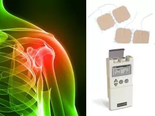
RCT- Non-operative Rx • 1. Painkillers and anti-inflammatory medications • 2. Physiotherapy • 3. Cortisone steroid injections • Reduces inflammation and control the pain. • Avoid repeated steroid injections in the presence of a tendon tear, as this may weaken the tendon further. • Outcome following Non-op Rx (MamanJBJS2009) • >50% FT and @8% PTRCTs progressed • 17% deterioration if <60 yrs, 54% if >60 yrs • Fatty infiltration results in increase tear size
RCT – Operative Rx • Single vs. Double row (DeHaan AM AJSM 2012) • Single-row repairs did not differ from the double-row repairs in functional outcome scores • Trend toward a lower retear rate in DR , although the data did not reach statistical significance • All arthroscopic vs. Mini-open repair (van der Zwaal P Arthroscopy 2013), (Kim SH Arthroscopy 2003) • Functional outcome, pain, range of motion, and complications do not significantly differ • Patients do attain the benefits of treatment somewhat sooner • Surgical outcome depended on the size of the tear, rather than the method of repair
Partial Thickness RCT • Articular side (PASTA) or bursal surface • O/E Like impingement, strength often reasonable • Pre-op diagnosis difficult, MRI inconclusive • Beware young patient with PTRCT, other aetiology than impingement • Initial conservative Rx appropriate
Partial Thickness RCT • The "50% rule“(Pedowotz RA ,Arthroscopy 2012) • Little scientific information is available to support the 50% rule • Significant PT tears need repair, not debridement (Weber OCNA, Arth 1999), (Kartus Arth 2006) • 1 in 5 (18%) re-op rate with debridement, progression to FT tears not uncommon • Acromioplasty and cuff debridement does not protect tear
Massive Cuff Tears • More common in older people, unusual under 60 years. • In patients with cuff degeneration • Disabling pain and weakness, pseudo paralysis • Marked atrophy and fatty infiltration poor clinical outcomes
Massive Cuff Tears - Rx • Non-op • Injection • Deltoid rehab prog • Operative • SAD, LHB tenotomy, Debridement • SS nerve ablation • Tendon transfers (Younger patient with irreparable RCT) • Reverse Shoulder Replacement
Shoulder Instability • Glenohumeral stabilisers • Static restraints • Glenohumeral ligaments (below) • Glenoid labrum (below) • Articular congruity and version • Negative intraarticular pressure • Dynamic restraints • Rotator cuff muscles • the primary biomechanical role of the rotator cuff is stabilizing the glenohumeral joint by compressing the humeral head against the glenoid • Biceps • Periscapular muscles
Dislocation Categories 1.Traumatic Dislocation ABankart lesion is the most common injury but other injuries can occur • HAGL tear • Bony Bankart • Hill-Sachs lesion 2. Atraumatic dislocation associated with joint laxity 3. Positional Non-traumatic dislocation 'abnormal muscle patterning' (party tricks)
Clinical tests – Instability • Anterior instability - anterior Apprehension - Jobe Relocation (Fulcrum Test)- anterior Drawer Test - anterior Load and Shift • Posteriorinstability - posterior Apprehension test - posterior Drawer Test - posterior Load and Shift • Inferior Laxity - Sulcus Sign
Instability Investigations • Plain X-ray • Initial imaging • MR arthrogram • Imaging modality of choice to evaluate the labrum • Associated ST lesions • CT arthrogram • Detection of bony injuries like glenoid rim # or HAGL • Also capsuloligamentous lesions
Shoulder Instability Rx • Non-op Rx • What position of immobilisation? ER or IR • Liavaag S JBJS(Am) 2011 • Immobilization in ER does not reduce the rate of recurrence for patients with first-time traumatic anterior shoulder dislocation • Physiotherapy - to train the shoulder muscles to control the shoulder correctly and prevent further instability • Operative Rx • A number of procedures are available depending on the causes and findings on investigations. • arthroscopic Procedures • Open Shoulder Procedures • Latarjet procedure for glenoid bone loss or • open capsular repair for HAGL lesions
Shoulder Instability Rx • Arthroscopic Stabilisation (Bankart Repair) • Repairing the over stretched or torn labrum and capsule • Latarjet-Bristow Procedure (transfer of the coracoid with it's attached muscles to the deficient area over the front of the glenoid) • Success due to the ‘triple effect’ described by Patte. • 1) Increase the glenoid contact surface area; • 2) The conjoint tendonreinforces theinferiorsubscapularis and anteroinferiorcapsule(Sling effect) • 3) Capsular repair
Frozen shoulder • Frozen Shoulder is an extremely painful condition • Often starts acutely, but may be triggered by a mild injury to the shoulder. • Frozen shoulder may be associated with diabetes, high cholestrol, heart disease and Dupuytrens contracture. • The capsule and its ligaments becomes inflamed, swollen, red and contracted. The normal elasticity is lost
Frozen Shoulder-Stages • Three stages • 1)Freezing phase: • Pain increases with movement and is often worse at night. • Progressive loss of motion with increasing pain. • Lasts approx. 2 to 9 months. • 2)Frozen phase: • Pain begins to diminish, • ROMmuch more limited • This stage may last 4 to 12 months. • 3)Thawing phase: • May begin to resolve. • Gradual restoration of motion over the next 12 to 42 months
Frozen shoulder Rx • Improve over 2-4 years after onset. • Painful &stiff shoulder generally require treatment. Rx modalities • Physiotherapy • Analgesics &Anti-inflammatories • Injections - reduce inflammation and provide pain relief • Hydrodilatation Procedure • MUA & Injection • Surgery - Arthroscopic Capsular Release . Intensive physiotherapy is essential after the surgery.
Frozen Shoulder Capsular Release • Over 80% success and the freedom from pain is quicker than MUA. • Diagnose other associated pathologies • Capsular release is safer and more effective than MUA for people who have developed a resistant stiff (frozen) shoulder after injury, trauma or fractures, as well as for diabetics.