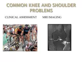
Comprehensive Guide to Common Shoulder Disorders
Understand basic shoulder anatomy, impingement syndrome, rotator cuff pathology, and more. Learn about shoulder muscles, bursa, and diagnostic tests. Discover treatment options, including conservative and operative approaches.

Comprehensive Guide to Common Shoulder Disorders
E N D
Presentation Transcript
Common Shoulder Disorders Abdulaziz Al-Ahaideb د عبدالعزيز الأحيدب MBBS, FRCS(C)
Basic shoulder anatomy • Impingement syndrome • Rotator cuff pathology • Adhesive capsulitis • Acromioclavicular pathology • Recurrent shoulder dislocations
Shoulder Anatomy • The greatest range of motion body.
Shoulder Anatomy:Bony Anatomy • Humerus • Scapula • Glenoid • Acromion • Coracoid • Scapular body • Clavicle • Sternum
Bones • Humerus. • Scapula (acromin): Type I : flat Type II: curved Type III: hooked • Clavicle
Joints • Glenohumeral joint: the main joint • Acromioclavicular (AC) joint • Sternoclavicular (SC) joint • Scapulothoracic joint
Glenohumeral Joint • Most common dislocated joint • Lacks bony stability • Composed of: • Fibrous capsule • Ligaments • Surrounding muscles • Glenoid labrum
Shoulder Anatomy:Rotator Cuff Muscles • Depress humeral head against glenoid
Shoulder anatomy:Rotator cuff muscles • Supraspinatus: • Abduction • Infraspinatus: • External rotation • Teres Minor: • External rotation • Subscapularis: • Internal rotation
Muscles • Deltoid: • largest, strongest muscle of the shoulder.
Shoulder Anatomy:Other Musculature • Pectoralis major, latissimus dorsi, biceps • Rhomboids, trapezius, levator scapulae, serratus anterior
Subacromial bursa • Between the acromion and the rotator cuff tendons. • Protects the acromion and the rotator cuff from grinding against each other.
Impingement Syndrome • Describes a condition in which the supraspinatus and bursa are pinched as they pass between the head of humerus (greater tuberosity) and the lateral aspect of the acromion
Risk factors • Age: over 40 years • Overhead activities • Bursitis and supraspinatus tendinitis • Acromial shape: type II & III acromion • AC arthritis or AC joint osteophytes may result in impingement and mechanical irritation to the rotator cuff tendons
Risk factors • Age (middle and older age; 40-85y) • Activity (overhead e.g. lifting, swimming, tennis). • Acromial shape. • Posterior shoulder capsule stiffness. • Rotator cuff weakness.
Symptoms • Pain in the acromial area when the arm is flexed and internally rotated Inability to use the overhead position. • The pain may result from subacromial bursitis or rotator cuff tendinitis • Pain when sleeping on the affected side.. • Pain will often become worse at night, as the subacromial bursa becomes hyperemic after a day of activity • Decreased range of motion especially abduction • Weakness
Differential diagnosis • Rotator cuff tears • Calcific tendinitis • Biceps tendinitis • Cervical radiculopathy • Acromioclavicular arthritis • Glenohumeral instability • Degeneration of the glenohumeral joint.
Physical examination • Atrophy of rotator cuff muscles. • Decreased range of motion (esp. internal rotation & adduction) • Weakness in flexion and external rotation. • Pain on resisted abduction and external rotation. • Pain on “impingement tests”..
Impingement tests • Neer’s impingement test: passive elevation of the internally rotated arm in the sagittal plane (shoulder forward flexion). • Hawkins’ impingement test: with the elbow flexed to 90 degrees, the shoulder passively flexed to 90 degrees and internally rotated.
Neer’s test Hawkins test
Radiological findings • Plain X-rays: • Acromial spurs • AC joint osteophytes • Subacromial sclerosis • Greater tuberosity cyst • MRI: • To confirm the diagnosis and rule out rotator cuff tear
Type of acromion: I flat II round III hooked Supraspinatous outlet view
Management • Conservative treatment: • Always start with it • Operative: • Indicated when conservative measures fail
Conservative treatment • Avoid painful and overhead activities • Physiotherapy: • Stretching and range of motion exercises • Strengthening exercises • NSAIDs • Steroid injection into the subacromial space
Operative treatment • The goal of surgery is to remove the impingement and create more subacromial space for the rotator cuff • Indicated if there is no improvement after 6 months of conservative treatment • The anterolateral edge of the acromion is removed • Open (called: Acromioplasty) or arthroscopic technique (called subacromial decompression) • Success rate 70-90%
Rotator cuff muscles • Supraspinatus: • Initiation of abduction + external rotation • Infraspinatus: • External rotation • Subscapularis: • Internal rotation • Teres Minor: • Internal rotation
Cont” Function of rotator cuff muscles • Keep the humeral head centered on the glenoid regardless of the arm’s position in space. • Generally work to depress the humeral head while powerful deltoid contracts
Causes of rotator cuff tears • Intrinsic factors: • Vascular • Degenerative ( age-related) • Extrinsic factors: • Impingement • Acromial spurs • AC joint osteophytes • Repetitive use • Traumatic (e.g. a fall or trying to catch or lift a heavy object)
Diagnosis • History • Physical examination • X-rays • MRI
Wide spectrum • Partial • Complete • Small • Large • Massive (irreparable)
Treatment • Degenerative type: (always start with non-operative) • Rest • Physio • NSAIDs • Steroid injection • If no improvement of 6 months, surgical repair (open or arthroscopic) is indicated • Traumatic type: (acute surgical repair)
If not treated chronic pain and loss of motion and with time becomes irreparable rotator cuff arthropathy • Complications of surgery: not improving, stiffness
Adhesive Capsulitis • Also called “frozen shoulder” • It is characterized by pain and restriction of all movements of the shoulder (global stiffness) • Usually self limiting (typically begins gradually, worsens over time and then resolves but may take >2 years to resolve) • 10 % is bilateral
Risk factors: • DM (esp. insulin dependent) • Hypo and Hyperthyroidism • Following injury or surgery to the shoulder • High cholestrol
Diagnosis: • Mainly clinical • X-rays and MRI to rule out other pathologies • Stages: • Pain (freezing stage) • Stiffness (frozen stage) • Resolution (thawing stage)
Adhesive Capsulitis Treatment • Resolves if untreated over 2-4 years • Physiotherapy • Pain and anti-inflammatory medications • Steroid injections • Manipulation under anesthesia • Arthroscopic capsular release
Acromioclavicular Pathology • The AC joint is different from joints like the knee or ankle, because it doesn't need to move very much. The AC joint only needs to be flexible enough for the shoulder to move freely. The AC joint just shifts a bit as the shoulder moves.
Causes of AC Arthritis • Degenerative osteoarthritis.( wear and tear in old aged people) • Rheumatoid Arthritis . • Gouty Arthritis. • Septic Arthritis. • Atraumatic distal claivcle osteolysis in weight lifters.
AC arthritis • Arthritis is a condition characterized by loss of cartilage in the joint, which is essentially wear and tear of the smooth cartilage which allows the bones to move smoothly. • Motions which aggrevate arthritis at the AC joint include reaching across the body toward the other arm.
Causes of AC osteoarthritis • Degenerative osteoarthritis.( wear and tear in old aged people) • Rheumatoid Arthritis • Gouty Arthritis • Septic Arthritis • Atraumatic osteolysis in weight lifters. ( result of repeated movements that wear away the cartilage surface found at the acromioclavicular joint) • Post-traumatic osteolysis of lateral end of clavicle.( like dislocation or a fracture)
Signs and Symptoms Pain , which worsens with movement and progressively worsens.( the patient may suffer a night pain which is a sign of arthritis) It is commonly associated with impingement syndrome Diagnosis: Clinical and by x-rays
AC osteoarthritis Non-surgical Treatment • Rest , avoid weightlifting and push-ups • Pain medications and NSAID to reduce pain and inflammation