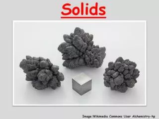
Bioviscoelastic Solids
1.08k likes | 1.29k Vues
Bioviscoelastic Solids. Prof. Ming-Shaung Ju Dept. of Mechanical Engineering NCKU. 7.1 Introduction. Soft tissues: muscles, blood vessel, ligament, tendon, cartilage, nerve Elastic materials: abductin, resilin, elastin, collagen

Bioviscoelastic Solids
E N D
Presentation Transcript
Bioviscoelastic Solids Prof. Ming-Shaung Ju Dept. of Mechanical Engineering NCKU
7.1 Introduction • Soft tissues: muscles, blood vessel, ligament, tendon, cartilage, nerve • Elastic materials: abductin, resilin, elastin, collagen • Thermodynamics of elastic deformation: two sources of elasticity • Constitutive equations of soft tissues • Uni-axial tension & quasilinear viscoelasticity • Biaxial loading
7.1 Introduction (cont’d) • 3D stress & strain in large deformation, pseudo-strain energy function • Example: skin • General viscoelastic relation • Computing strain from known stress
7-2 Some Elastic Materials • Actin 肌動蛋白 • Elastin 彈性蛋白 • Collagen 膠原蛋白 • Aldehyde 醛 • Resilin 彈性蛋白 • Abductin 外展蛋白
7.2.1 Actin • In all muscles, leukocytes, red blood cells, endothelial cells & many other • Strength of a single actin filament (Kishino & Yanagida, 1988) • Single actin filament (~7nm f) labelled with phalloidin-tetramethyl-rhodamine • Two microneedles connected to micromanipulators; one very flexible & the other stiff; • Needles coated with monometric myosin to increase affinity with actin
Actin (cont’d) • Stiff needle was pulled until actin broke; force was calculated from bending of flexible needle • For actin filaments of length 4 to 32 um, tensile force was 108 +/- 5 pN (n=61) without breaking, comparable to single unit in muscle during isometric contraction • Tensile strength 108pN*4/p72(nm)2=2.2x106N/m2 or 2.2MPa
7.2.2 Elastin (彈性蛋白) • Most linearly elastic biosolid material • Cylindrical specimen of elastin subjected to uni-axial load
Ligamentum nuchae (項韌帶) denature(變質) Tensile strain: change of length / initial length Stress: load / initial cross sectional area (at zero stress) Almost linear with small hysteresis Elastic limit up to l =1.6
Elastin (cont’d) • Protein found in vertebrates • Thin strands in skin & in areolar(蜂窩) connective tissue; walls of arteries & veins, especially near heart; prominent component of lung tissue • Ligamentum nachae is almost pure elastin; • small amount of collagen can be denatured by heating to 66◦C +. • Note the process does not change properties of elastin • Provide elasticity to arteries & lung parenchyma tissue; keep skin tissue smooth;
7.2.3 Incomplete fixation of elastin in Aldehyde (醛) • Fixation is commonly used in histology studies. When the tissue has elastin in it artifacts may induced by elastic recovery • Tissue usually fixed by formalin, formaldehyde(蟻醛), or glutaraldehyde then embedded, sectioned & stained. • If elastin specimen is stretched under tension & soaked in these agents, on release of tension it does not return to unstretched length (40%-70% recovery) but remain elastically
Elastic recovery of elastin after fixation in formalin & glutaraldehyde Note: elastic recovery occurs at all stretch ratios, it is not fixed !
If a tissue is fixed in a state of tension, e.g., inflated lung, or a disintended artery, & • sectioned under no load, residual stress in elastin fibers will be released, • length of elastic fibers will be shortened to length at zero stress state. • Fixed part of tissue will be buckled by shortening of elastin
(a) • Lung parenchyma of a spider monkey, fixed in glutaraldehyde & embedded in wax. Note: tissue was allowed to shrink in a stress-free state. (b) Same tissue embedded in celloidin, a hard plastic. Tissue is not allow to shrink. (b) Pulmonary alveoli 肺泡
Winkle appearance of most photomicrographs of lung tissue is artifact caused by unsuspected elastic recovery of elastin fibers!
7.2.4 The Elastin Molecule • Molecular structure of tropoelastin been sequenced; tropoelastin formed intracellularly & cross-linked extracellulary. • Poly (V PG VG), poly (V PG F GV G AG), poly (VPGG) on g-irradiation cross-linking are elastic • These polypeptides will self-assembly into more ordered molecular assemblies on raising temperature.
Elastin Molecule (cont’d) • Sources of elasticity • Decrease of entropy • Increase of internal energy • Entropy theory: liberation mechanism or rocking of peptides segment contributed to entropy S =k ln W • Self assembly mechanism has a critical temperature of 25。C • From lung tissue of rat Debes & Fung suggests that inverse temperature transition phenomenon may not a major mechanism for whole elastin
7.2.5 Resilin & Abductin • Resilin: similar to elastin in mechanical behavior, different in chemical composition • Protein in arthropods (節足動物), hard when dried, soft & rubbery in natural state 50-60% water. • Within stretch ratio l=1-2, E~1.8x10⁷ dyn/cm2 or 1.8 MPa; G~0.6MPa • Insects uses resilin as elastic joints for wings; fleas & locusts use resilin at base of hind legs as catapults in jumping. • Abductin: in scallops’ hinges to open valves • About same elastic moduli as elastin
7.2.6 Elasticity due to entropy & Internal Energy Changes • Elastin, resilin & abductin, long flexible molecules jointed together by cross-linking to form 3D networks • Molecules are convolute and thermal energy keeps them in constant thermal motion; • Configurations (entropy) change with strain; from entropy change elastic stress appears.
Crystalline materials derive elastic stress from changes in internal energy, i.e., elastic modulus related to strain of crystal lattice. • Eq.(1) does not apply to crystalline materials, fibers whose elasticity comes partly from internal energy changes & partly from entropy changes. • Most biological materials that can sustain finite strain have rubbery elasticity but not all. • Hair can be stretched to 1.7 times & spring back but this is due to keratin has two crystalline forms a & b helices; when stretched some a helices change to b helices.
7.2.8 Crystallization due to strain • Raw rubber can be stretched several times its length & held extended; stress relaxation is almost complete • Stress relaxation of rubber is due to crystallization. Stretching extends molecules so they run parallel to each other and crystallize. Heating disrupts the crystalline structure. • Silk is crystallized under a high shear strain rate and emerge as a fiber. Composition: fibroin(繭絲蛋白) & sericin(絲蛋白); sericin is gummy & dissolvable in warm water, • Young’s modulus of silk: 104MPa, broke at l=1.2
7.3 Collagen • Basic structural element for soft & hard tissues in animals, providing mechanical integrity & strength to our bodies. • Important to man as steel to civilization. • Main load carrying element in blood vessels, skin, tendons, cornea, sclera(鞏膜), bone, fascia(筋膜), dura mater(硬腦膜), uterian cervix(子宮頸). • Collagen molecules, how they wind into fibrils and how fibrils organized into fibers, fibers into tissues.
In each stage of organization, new features of mechanical properties are acquired. • In physiology & biomechanics, major attention focused on organ & tissue level. • Relationship between function & morphology of collagen in different organs.
7.3.1 Collagen Molecules • A protein containing sizable domains of triplet-helical conformation and functioning as supporting elements in an extra-cellular matrix. • Arrangement of amino acids: every 3rd residue glycine(甘膠酸), proline & OH-proline follow each other; 3 residues per turn; left hand helices • Chains coiled follow a right-hand twist w. a pitch of 8.6 nm; 3 helical chains arranged w. slight displacement longitudinally; 0.291 nm (-110 deg); distance between each 3rd glycine 0.873 nm
Types of collagens • 12 types of collagens has been identified • a1(I): a chain of type I collagen
7.3.2 Aggregate Structure • Relation of function and structure of collagen aggregates (Miller, 1988) • Fiber-forming collagens
A: I, II, III, V, K B: IV basement membrane C: VI: placental villi(胎盤絨毛) D: VII: placental membrane
Distribution of collagens in human body • Type I, ubiquitous, can be isolated from any tissue or organ, e.g., bone, dermis, placental membrane & tendon • Type II, mainly in hyaline cartilage and cartilage-like tissues such as nucleus pulposus of vertebral body & body of eyes. • Type III, constitutants of dermis, blood vessel walls & other more distensible connective tissues; ubiquitous too. • Type V, relative minor but has distribution like type I • Type K (XI), like Type II, mainly, in cartilage • Type IX & X, minor constituents of hyaline cartilages; short-chain collagens.
7.3.3 Collagen fibrils & fibers • Fiber-forming collagen molecules: collection of tropocollagen molecules forms a collagen fibril; • Fibril appears cross-striated (EM graphs), examples: tendon & skin • Period of striation D = 64 nm (native) or 68 nm (moisture) • A gap of 0.6D is left between ends of successive molecules. The gap appears as the lighter part of the striation. • Current view, not perfectly parallel, bent, varying spacing
EM graphs of tendon & skin skin tendon X 24,000
Model of organization of molecules Quarter-stagger Length of each molecule/ D = 4.4
Diameter of fibril 20-40 nm, depends on animal species & tissue. • Bundles of fibrils form fibers, diameter 0.2 to 12 um. Fibers are colorless under light microscope and birefringent in polarized light; • In tendon as long as tendon itself, in connective tissues length varies considerably; • Packaging of collagen fibers has many hierarchies: parallel-fiber for tendon, (Fig.7.3:6) • Fibers assembled into fascicles & enclosed in a sheath of reticular membrane to form a tendon.
7.3.4 Wavy Course of Fibers • Rat’s tail tendon in polarized light microscope observation light & dark pattern period 100 um: waviness of collagen fiber in fascicle • When tendon is stretched waviness of crimped fibers decreases and wave shape is planar. • Wave parameters (Table 7.3:2) • When stretched bending angle q0 decreases • Fiber diameter is age dependent. For rat, it increases from 100 to 500 nm as rat ages!
Basic mechanical unit of tendon is bent collagen fibers. • Q: Are the fibers intrinsically bent because of some fine structural features of the fibrils ? • A: curvature of fibers might be caused by shrinking of non-collagen components or “ground substance” of tendon, i.e., curvature is due to buckling of fibers. • Integrity of ground substance is important for mechanical integrity of tendon. • Enzymatic digestion directed at non-collagen components changes mechanical properties of tendon. • Buckling model (Dale & Baer, 1974): hyaluronic acid(玻尿酸) may be responsible for the buckling of collagen fibers.
In some tissues, elastin & collagen together form a composite material. Straight elastic fibers are attached to bent collagen fibers. Not for pulmonary alveolar.
7.3.5 Ground Substance (stroma) • Collagen integrated with cells & intercellular subatance • Dense connective tissues: fibrocytes, fibers of collagen, elastin, reticulin, hydrophilic gel (ground substance) • Loose connective tissues have more amount of ground substance than dense connective tissues • Composition: mucopolysaccharides(黏多醣) or glycosaminoglycans(葡萄糖胺), tissue fluid; • Hydration of collagen is an important problem in biomechanics!
7.3.6 Structure of Collagenous Tissues • Parallel fibers: tendons & ligaments; 2D & 3D networks: skins & blood vessels, intestinal mucosa & female genital tracts. • Tendon functions transmit tension & parallel-fiber; • Ligament similar but less regular, curved & oblique to motion direction; most collagenous, ligamenta flava of spine & ligamentum nuchae mostly elastin. • Insertion of ligaments to bones, gradual transition from ligament to bones; rows of fibrocytes, chondrocytes, groups of osteocytes;
Tendon inserts one end to main fibrous layer of periosteum. The other end to muscle, collagenous fibrils bound to plasma membranes & collagen fibers. • In fasciae(筋膜) & aponeuroses(腱膜), parallel fibers spread into sheets. • Others include: diaphragm, periosteum, perichondrium, membrana fibrosa of capsules, dura matter, sclera, fasciae, organ capsules. • Collagen fibers in skin – 3D networks of fibrils, woven into rhombic parallelgram pattern. In dry dermis, 75% collagen & 4% elastin. • Collagen structure in blood vessel (Sec 8.2) • Female genital tract muscle organ, smooth muscle cells in circular & spiral pattern; in uterus 30-40%v muscle, in cervix 10%v muscle, other connective tissues,
7.3.7 The Stress-Strain Relationship • Nonlinear load-elongation • Hysteresis & stress relaxation • Cyclic loading variation
Typical load-elongation for rabbit limb tendon • O-A toe: exponential increase • physiological region, normal • A-B linear, strength of tendon • B-C nonlinear • C 50-100MPa, maximum load • Rupture elongation 10-15 % • Slope tan a: elastic stiffness
韌帶機械特性 Toe