Download
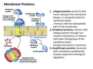
1 / 35
350 likes | 516 Vues
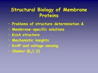
Bioinformatics and Molecular Modeling studies of Membrane Proteins. Shiva Amiri. Problem : Difficult to obtain high resolution crystallographic images of membrane proteins. Structure Determination. Unwin et.al , Nature , 26 June 2003. Getting there?.

E N D
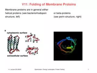
Bioinformatics and Molecular Modeling studies of Membrane Proteins Shiva Amiri
Problem: Difficult to obtain high resolution crystallographic images of membrane proteins StructureDetermination Unwin et.al, Nature, 26 June 2003
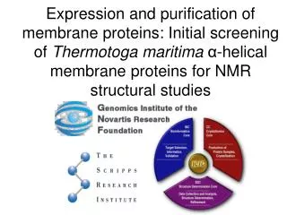
Getting there? • Some success using cryo-electron microscopy coupled with Fourier Transforms, i.e. Unwin’s 4 Å image of the TM region. • But still no full structure Unwin et al., Nature, 26 June 2003
My Project • To design structure determination software for Ligand Gated Ion Channels (LGICs) i.e. nAChR, GABAA and GABAC receptors, 5HT3 receptor, Glycine receptor
Main focus so far: The α-7 Nicotinic Acetylcholine Receptor (nAChR) • cationic channel • homopentamer • four transmembrane regions (M1-M4)
Homology modeling – Transmembrane domain Transmembrane Domain Alignment
Homology modeling – Transmembrane domain • The homology model of the TM region with the Torpedo Marmorata structure (PDB: 1OED - 4 Å) and the Chick α-7 sequence using MODELLER
Homology Modeling – Ligand Binding domain Ligand Binding Domain Alignment
Homology Modeling – Ligand Binding domain • The homology model of the LBD with Acetylcholine Binding Protein (AChBP) as the structure (PDB: 1I9B – 2.7 Å) and the Chick α-7 sequence using MODELLER
The software … • Combining the Transmembrane domain with the Ligand Binding domain • Failed first attempt: Minimizing distances between target residues in the LBD and the TM domains - 6 degrees of freedom (rotations and translations on all three axes) - Models were not straight
Second attempt … New algorithm: straighten each domain with respect to the z-axis • Align each domain onto the z-axis z-axis
Theta (angle of rotation) z-axis Second Attemptcontinued… • Rotate and translate about z-axis - angle of rotation and steps of translations are user- defined
At each rotation and for each translation the Unwin distance, the Termini distance the number of bad contacts is calculated
Scoring Functions • Unwin Distance – the distance between residues from the TM domain and the LB domain that are meant to come into close proximity
Scoring Functions continued … • Termini Distance – the distance between the C-terminus of the LB domain and the N-terminus of the TM domain
Scoring Functions continued… • Bad Contacts – Number of residues that are closer than a certain cut-off distance (user-defined), currently set to 5 Å
Plots of Scoring Functions Unwin Distance Termini Distance Bad contacts
Linear Combinations of Scoring Functions Unwin + Termini Unwin + Termini + Bad contacts
Choosing the Best Model • Model chosen based on scoring function data • Once a good model was decided on, energy minimization using GROMACS was carried out to ensure the electrochemical legitimacy of the model
Gaussian Network Model (GNM) Analysis • A course-grained model to approximate molecular motions of proteins • Current code cannot allocate memory for the 1660 residues of the α-7 nAChR • Analysis has been done using the TM domain and the LB domain separately • GNM was also run on one subunit of the model • B-values generally in agreement with crystallographic data but modeled structures are difficult to analyze using present code
CONCOORD Analysis • Generates protein conformations around a given structure based on distance restrictions • Principal Component Analysis (PCA) is applied on the 500 resulting structures from CONCOORD • First eigenvector shows opening and closing of the pore as the subunits rotate
Eigenvector Plot Covariance lines
Future work… Summary • Software designed to determine structure of LGICs • Structure of α-7 nAChR • Used various methods (GNM, CONCOORD) to look at possible motions using the hypothesized structure • Looking at the hydrophobic girdle (M2) of LGICs to study patterns of conservation and the behaviour of these residues during gating • Further verification and analysis of models • Other models of LGICs
Thanks to: Prof. Mark S.P. Sansom Sundeep Deol Dr. Phil Biggin Yalini Pathy Dr. Kaihsu Tai Jonathan Cuthbertson Dr. Paul Barrett Pete Bond Jeff Campbell Dr. Alessandro Grotessi Katherine Cox Dr. Daniele Bemporad Jennifer Johnston Dr. Jorge Pikunic Robert D’Rozario Dr. Shozeb Haider Loredana Vaccaro Dr. Andy Hung John Holyoake Dr. Bing Wu Tony Ivetac Oliver Beckstein Sylvanna Ho Syma Khalid Samantha Kaye Zara Sands George Patargias
Hydrophobic girdle M2 alignment
Rotate and translate about z-axis - angle of rotation and steps of translations are user- defined