Download
1 / 79
790 likes | 814 Vues
Explore the mechanisms of passive diffusion, facilitated diffusion, active transport, specialized membrane pores, ionophore antibiotics, and more. Understand processes like symport, antiport, and secondary active transport driven by ion gradients. Learn about porins, pore-forming toxins, and amphipathic helices forming transmembrane channels.

E N D
Membrane Proteins and Transport BL4010 11.28.06
Outline • Passive Diffusion • Facilitated Diffusion • Active Transport • Transport Driven by ATP, light, etc. • Specialized Membrane Pores • Ionophore Antibiotics
http://www.stolaf.edu/people/giannini/flashanimat/transport/osmosis.swfhttp://www.stolaf.edu/people/giannini/flashanimat/transport/osmosis.swf
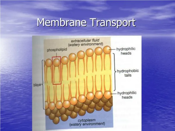
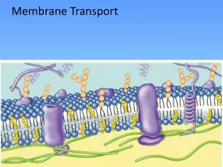
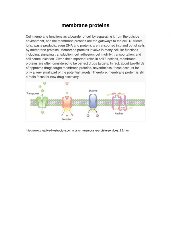
Passive Diffusion No special proteins needed • Transported species simply moves down its concentration gradient - from high [c] to low [c] • High permeability coefficients usually mean that passive diffusion is not the whole story
Facilitated Diffusion G negative, but proteins assist • Solutes only move in the thermodynamically favored direction • But proteins may "facilitate" transport, increasing the rates of transport • Two important distinguising features: • solute flows only in the favored direction • transport displays saturation kinetics
Facilitated DiffusionBeware the cartoon world! • Uniport • http://www.stolaf.edu/people/giannini/flashanimat/transport/caryprot.swf • Gated channel • http://www.stolaf.edu/people/giannini/flashanimat/transport/channel.swf • Symport • http://www.stolaf.edu/people/giannini/flashanimat/transport/symport2.swf
Active Transport Systems Energy input drives transport • Some transport must occur such that solutes flow against thermodynamic potential • Energy input drives transport • Energy source and transport machinery are "coupled" • Energy source may be ATP, light or a concentration gradient
Secondary Active Transport Transport processes driven by ion gradients • Many amino acids and sugars are accumulated by cells in transport processes driven by ion gradients
Secondary Active Transport • Symport - ion and the amino acid or sugar are transported in the same direction across the membrane • Antiport - ion and transported species move in opposite directions • Several examples are described in Table 10.2
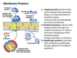
Porins Found both in Gram-negative bacteria and in mitochondrial outer membrane • Porins are pore-forming proteins - 30-50 kD • General or specific - exclusion limits 600-6000 • Most arrange in membrane as trimers • High homology between various porins • Porin from Rhodobacter capsulatus has 16-stranded beta barrel that traverses the membrane to form the pore (with eyelet!)
Why Beta Sheets? for membrane proteins?? • Genetic economy • Alpha helix requires 21-25 residues per transmembrane strand • Beta-strand requires only 9-11 residues per transmembrane strand • Thus, with beta strands , a given amount of genetic material can make a larger number of trans-membrane segments
The Pore-Forming Toxins • Lethal molecules produced by many organisms • They insert themselves into the host cell plasma membrane • They kill by collapsing ion gradients, facilitating entry by toxic agents, or introducing a harmful catalytic activity
Colicins • Produced by E. coli • Inhibit growth of other bacteria (even other strains of E. coli) • Single colicin molecule can kill a host! • Three domains: translocation (T), receptor-binding (R), and channel-forming (C)
colicin Ia, 210 Å spans periplasmic space of gram negative bacteria. blue=receptor binding (o.m.) violet=C-domain forms channel on i.m. green=hydrophobic red=translocon
Clues to Channel Formation • C-domain: 10-helix bundle, with H8 and H9 forming a hydrophobic hairpin • Other helices amphipathic (Fig. 10.30) • H8 and H9 insert, with others splayed on the membrane surface • A transmembrane potential causes the amphipathic helices to insert!
Other Pore-Forming Toxins • Delta endotoxin also possesses a helix-bundle and may work the same way • There are other mechanisms at work in other toxins • Hemolysin from Staphylococcus aureus forms a symmetrical pore • Aerolysin may form a heptameric pore - with each monomer providing 3 beta strands to a membrane-spanning barrel
Hemolysin allows Ca2+ influx, kills blood cells (Staph. aureus)
Amphiphilic Helicesform Transmembrane Ion Channels • Many natural peptides form oligomeric transmembrane channels • The peptides form amphiphilic -helices • Aggregates of these helices form channels that have a hydrophobic surface and a polar center • Melittin (bee venom), magainins (frogs) and cecropin (from cecropia moths) are examples
Melittin • Bee venom • 26 amino acids • monomer in membrane until potential is applied then tetrameric chlorid channel • causes pores to form in nocireceptors (pain) • stimulates the nerve, pain response, resetting of nerve causes deoligomerization but re-established with next resting potential
Amphipathic Helices • Melittin - bee venom toxin - 26 residues • Cecropin A - cecropia moths - 37 residues • Magainin 2 amide - frogs - 23 residues • See Figure 10.35 to appreciate helical wheel presentation of the amphipathic helix
The Magainin Peptides • Discovered by Michael Zasloff • He noticed that incisions on Xenopus laevis (African clawed frog) healed without infection, even in bacteria-filled aquarium water • He deduced that the frogs produced a substance that protected them from infection!
The Cecropins • Produced by Hyalophora cecropia Induced when the moth is challenged by bacterial infections • These peptides are thought to form helical aggregates in membranes, creating an ion channel in the center of the aggregate
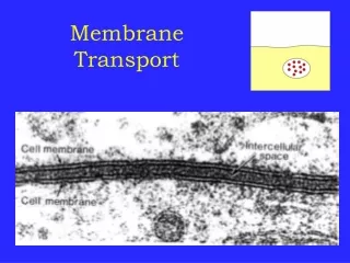
Gap Junctions Vital connections for animal cells • Provide metabolic connections • Provide a means of chemical transfer • Provide a means of communication • Permit large number of cells to act in synchrony
Gap Junctions • Hexameric arrays of a single 32 kD protein • Subunits are tilted with respect to central axis • Pore in center can be opened or closed by the tilting of the subunits, e.g. as response to stress
Ionophore Antibiotics Mobile carrier or pore (channel) • How to distinguish? Temperature! • Pores will not be greatly affected by temperature, so transport rates are approximately constant over large temperature ranges • Carriers depend on the fluidity of the membrane, so transport rates are highly sensitive to temperature, especially near the phase transition of the membrane lipids
Valinomycin A classic mobile carrier • A depsipeptide - a molecule with both peptide and ester bonds • Valinomycin is a dodecadepsipeptide • The structure places several carbonyl oxygens in the center of the ring structure • Potassium and other ions coordinate the oxygens • Valinomycin-potassium complex diffuses freely and rapid across membranes
Selectivity of Valinomycin Why? • K + and Rb + bind tightly, but affinities for Na + and Li + are about a thousand-fold lower • Radius of the ions is one consideration • Hydration is another it "costs more" energetically to desolvate Na+ and Li+ than K+
Gramicidin A classic channel ionophore • Linear 15-residue peptide - alternating D & L • Structure in organic solvents is double helical • Structure in water is end-to-end helical dimer • Unusual helix - 6.3 residues per turn with a central hole - 0.4 nm or 4 A diameter • Ions migrate through the central pore
The Sodium Pump aka Na+/K+-ATPase • Large protein - 120 kD and 35 kD subunits • Maintains intracellular low Na+ and high K+ • Crucial for all organs, but especially for neural tissue and the brain • ATP hydrolysis drives 3Na+ out and 2K+ in • Alpha subunit has ten transmembrane helices with large cytoplasmic domain
The Na+/K+ ATPase • ATP hydrolysis drives 3Na+ out and 2K+ in