Download
1 / 32
330 likes | 540 Vues
Image Reconstruction and Image Priors. Vadim Soloviev, Josias Elisee, Tim Rudge, Simon Arridge. Munich April 24, 2009. TexPoint fonts used in EMF. Read the TexPoint manual before you delete this box.: A A A A. P4 University College London – Computer Science (UCL)

E N D
Image Reconstruction and Image Priors Vadim Soloviev, Josias Elisee, Tim Rudge, Simon Arridge Munich April 24, 2009 TexPoint fonts used in EMF. Read the TexPoint manual before you delete this box.: AAAA
P4 University College London – Computer Science (UCL) UCL has an annual turnover of £500M, academic and research staff totaling 4,000, and over 3,000 PhD research students. The department of Computer Science has over 50 academic staff with specialist groups involved in Imaging Science, Computer Graphics, BioInformatics.Intelligent Systems Networking, and Software Systems Engineering. CMIC In 2005, the Centre for Medical Imaging (CMIC) was formed jointly between Computer Science and the department of Medical Physics & BioEngineering to create a world class grouping combining excellence in medical imaging sciences withinnovative computational methodology, finding application in biomedical research and in healthcare. The research of the group focuses on detailed structural and functional analysis in neurosciences, imaging to guide interventions, image analysis in drug discovery, imaging in cardiology and imaging in oncology with a strong emphasis on e-science technologies. The Centre has very close links with the Faculty of Clinical Sciences, the Faculty of Life Sciences and associated Clinical Institutes, in particular the Institute of Neurology,the Institute of Child Health and the Centre for Neuroimaging Techniques (CNT), • Main tasks attributed to the organisation: The main tasks for P4 UCL are WP4 with some input into WP3, WP6 and WP7. We will contribute mathematical and computational techniques for the development of forward and inverse modeling in optical tomography in the diffuse regime particularly using priors, and to the simulation of new imaging devices and the analysis of clinical data.
Objective 4.1 : To develop FMT inversion utilizing XCT image priors without strong anatomy function correlations. Progress: • Developed structured priors orientating the reconstructed FMT images to have level sets parallel to those of XCT image • Developed information theoretic priors orientating the reconstructed FMT images to have maximum joint entropy with the XCT image • Initial tests on simulated 3D images of mouse from a realistic atlas Significant Results • Reconstructions in 3D depend only linearly on total number of pixels in reconstructed image and independent of the number of pixels in data. Deviations from Annex 1 • None Failure to meet critical objectives • Not Applicable Use of resources • No deviation from work
Choice of Prior Consider where and D a symmetric tensor By variation where Lagged_Diffusivity Gauss-Newton Method
Choice of Prior (2) 1st order Tikhonov Examples: Total Variation Now choose Where n is normal to level set of another image xref x xref
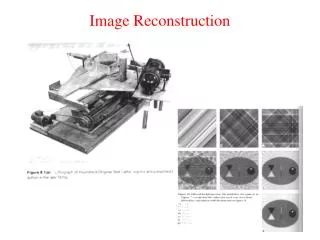
Example: Cylinder with inclusions Target object: cylinder with embedded inhomogeneities Radius: 25 mm, height: 50 mm Background: ma=0.01 mm-1, ms=1 mm-1 Red: Inclusions with increased absorption Blue: Inclusions with increased scattering Measurements: 80 source locations, 80 detector locations, arranged in 5 rings at elevations -20, -10, 0, +10, +20 Data: log amplitude and phase for source modulated at 100MHz Multiplicative Gaussian noise 0.5% FEM mesh: 83142 nodes, 444278 4-noded tetrahedra Reconstruction grid: 80x80x80 Cross sections through target for planes z=16, z=60 and y=40 ma ms
Reconstruction with flat TV prior ma ms Iso-surfaces Cross sections Reconstruction: Nonlinear conjugate gradients (50 iterations) with line search Prior: TV with hyperparameter t = 10-4 and smoothing parameter b = 0.1
Reconstruction with TV prior using correct structural information ma Edge prior ms Iso-surfaces Cross sections ma Reconstruction ms Iso-surfaces Cross sections
TV prior using undifferentiated structural information ma Edge prior ms Iso-surfaces Cross sections ma Reconstruction ms Iso-surfaces Cross sections
TV prior using partial structural information ma Edge prior ms Iso-surfaces Cross sections ma Reconstruction ms Iso-surfaces Cross sections
Reconstructions ma m’s
Objective 4.2 : To incorporate XCT image segmentation into the FMT Progress: • Developed segmentation of XCT based on anisotropic diffusion (Perona-Malik algorithm) • Developed hexahedral adaptive mesh generation from XCT images • Incorporated mesh reduction methods using public-domain software ISO2MESH • Developed Boundary Element (BEM) and hybrid Boundary-Finite Element (BEM-FEM) methods Significant Results • Reconstructions using FEM only for internal organs are much faster than using a complete FEM mesh Deviations from Annex 1 • None Failure to meet critical objectives • Not Applicable Use of resources • No deviation from work
Segmentation Requirements • construction of meshes for numerical modelling • construction of priors as required in Objective 4.1 • post-reconstruction object labelling and analysis
Objective 4.3 : To calculate spatially varying optical attenuation in tissues in-vivo. Progress: • Developed non-linear reconstruction method for attenuation making use of Louiville transformation from diffusion to Schrodinger equation. Significant Results • Reconstruction of attenuation from steady-state data is dependent on good estimates of spatially varying scatter. Deviations from Annex 1 • None Failure to meet critical objectives • Not Applicable Use of resources • No deviation from work
Objective 4.4 :To develop FMT inversion based on simultaneous XCT segmentation and classification Progress: • Developed combined reconstruction/segmentation method combining Gauss-Newton image reconstruction with fuzzy-kmeans image classification. • Developed fully hierarchical Bayesian framework Significant Results • Classification error less than 5% for simulated noisy data. Deviations from Annex 1 • None Failure to meet critical objectives • Not Applicable Use of resources • No deviation from work
Combined Reconstruction Classification y Cy Data Noise Statistics x Reconstruction Step Estimation Step Image x,Cx l,q Class Statistics Image Statistics Prior Update Step
Deliverables 4.1 Inversion algorithms • Deliverable 4.1 was created as an inversion code. Two versions were developed : • compiled C++ code using UCL Toast Libraries and OpenGL graphics • Matlab program using Mex version of TOAST libraries and Matlab Graphics • Individual installations on partner systems will be provided at the next project meeting.
Conclusions • FEM and BEM based solvers • Linear and non-linear reconstruction • Large Data Sets using Matrix-Free approach • Structural Priors incorporating image information, not dependent on segmentation • Statistical Priors based on information theory • Matlab based code available on web http://web4.cs.ucl.ac.uk/research/vis/toast/index.html