Image Processing Project Tomographic Image Reconstruction
Image Processing Project Tomographic Image Reconstruction. Introduction. This presentation will cover a brief description of tomographic imaging, image reconstruction methods, and design challenges. 1. Tomography. 2. Image Reconstruction. 3. Design Challenges. 4. Your Project.


Image Processing Project Tomographic Image Reconstruction
E N D
Presentation Transcript
Image Processing Project Tomographic Image Reconstruction Introduction
This presentation will cover a brief description of tomographic imaging, image reconstruction methods, and design challenges. 1. Tomography 2. Image Reconstruction 3. Design Challenges 4. Your Project
Background: Importance of Medical Imaging • There are an estimated 630,000 imaging procedures every week in the US. • The average radiologist has a case load around 35% higher than just 5 years ago. “Molecular imaging holds great promise for early detection and treatment of numerous diseases, for providing researchers with detailed information about cellular physiology and function, and for facilitating the goal of personalized medicine.” – NIH roadmap
Modern imaging modalities cover the EM spectrum and all scales of resolution. Organism Organs Tissue Cells Proteins Genes
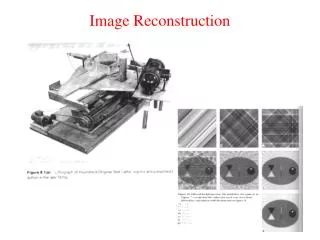
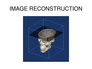
Tomography X-ray CT Images of the Human Abdomen X-ray CT Scanner
Tomography means to image using sections or slices, tomo is Greek for cut, graph means form (plot) an image. • Tomograms can be created using a variety of physical mechanisms • X-ray Attenuation • Nuclear Magnetic Resonance • Position-Electron Annihilation • Ultrasound Interactions • Modalities • X-ray Computed Tomography • Magnetic Resonance Imaging • Positron Emission Tomography • Ultrasound Interactions
To do Tomography, we need to have many projections from different angles. • Transforms the object we are imaging to a Sinogram
= density of 25 = density of 0 = density of 50
= density of 25 = density of 0 = density of 50
= density of 25 = density of 50 = density of 0
= density of 25 = density of 50
= density of 25 = density of 50
= density of 25 = density of 50 Our First Projection
= density of 25 = density of 50 Our First Projection
= density of 25 = density of 50 Our First Projection
= density of 25 = density of 50 Our First Projection
= density of 25 = density of 50 Our First Projection
Given all these projections, how do we reconstruct the tomogram? • Filtered Backprojection
Image Reconstruction Using Filtered Backprojection Filter Backprojection
Backprojection • Backprojection “smears” the data back.
Backprojection • Backprojection “smears” the data back.
Backprojection • Backprojection “smears” the data back. infinite number of projections
Filtered-Backprojection • If we filter the projections before backprojection we can recover the original object.
Filtering the projection • Filtering is a basic operation in signal processing. • Spatial or Temporal domain filtering (convolution) • Frequency domain filtering • Lets consider spatial domain filtering: input signal (projection) filtered signal filter kernel convolution operator
TPS: Think-Pair-Share • Match up the following Sinograms with their objects O1 S1 O? = S? O2 S2 O3 S3
Design Challenges • In cases where the projection is taken using x-rays, there is a risk associated. • x-rays absorbed by the body can cause damage to DNA directly or through the formation of free radicals. • The larger the number of projections and the longer the x-rays are on, the higher the dose delivered to the patient. • There is a fundamental tradeoff between dose, the number of projections, and the noise in the projections.
Basics of Radiation Biology • We are constantly exposed to naturally occurring radiation (radon, cosmic rays) • about 3 milli-Sieverts per year • there is some evidence that anti-oxidants, found in fruits and vegetables, can protect cells from free radical formation • A single chest x-ray is equivalent to about 10 days of natural exposure • A whole-body x-ray CT exam is equivalent to about 3 years of equivalent natural exposure
Radiation Monitoring • OSHA requires all those who work with radiation to be badged and monitored for dose.
Noise in X-rays • X-ray images are formed by counting the number of photons leaving the subject compared to those entering. • The count is a random variable that is Poisson distributed.