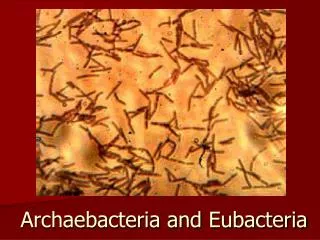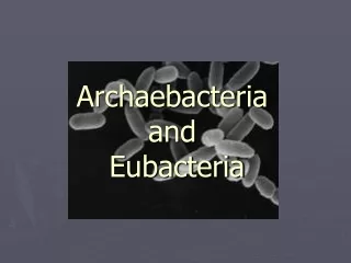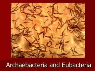
Archaebacteria and Eubacteria
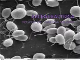
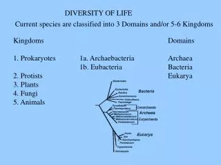
Archaebacteria and Eubacteria. Kingdom Archaebacteria. Prokaryotic Cell wall without peptidoglycan Heterotrophs Saprophytic or parasitic Asexual (binary fission) Live in extreme conditions. Eg. methanogens (animal gut), extreme thermophiles (Heat), extreme halophiles (Salty).
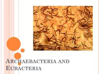

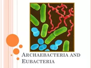
Archaebacteria and Eubacteria
E N D
Presentation Transcript
Kingdom Archaebacteria • Prokaryotic • Cell wall without peptidoglycan • Heterotrophs • Saprophytic or parasitic • Asexual (binary fission) • Live in extreme conditions Eg. methanogens (animal gut), extreme thermophiles (Heat), extreme halophiles (Salty)
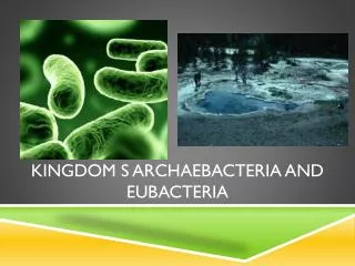
Kingdom Eubacteria 2 sub groups: Bacteria or Cyanobacteria (photosynthetic with chlorophyll in cytoplasm) • Prokaryotic • Cell wall contains peptidoglycan • Heterotrophs or autotrophs • Asexual (binary fission) • Saprophytic or parasitic • Lives anywhere
Bacteria are of immense importance because of their rapid growth, reproduction, and mutation rates, as well as, their ability to exist under adverse conditions. • The oldest fossils known, nearly 3.5 billion years old, are fossils of bacteria-like organisms.
Depending on the species, bacteria can be aerobic which means they require oxygen to live or • anaerobic which means oxygen is deadly to them. Green patches are green sulfur bacteria. The rust patches are colonies of purple non sulfurbacteria. The red patches are purple sulfur bacteria.
Methanogens These Archebacteria are anaerobes. They make methane (natural gas) as a waste product. They are found in swamp sediments, sewage, and in buried landfills. In the future, they could be used to produce methane as a byproduct of sewage treatment or landfill operation.
Halophiles These are salt-loving Archaebacteria that grow in places like the Great Salt Lake of Utah or salt ponds on the edge of San Francisco Bay. Large numbers of certain halophiles can turn these waters a dark pink. Pink halophiles contain a pigment very similar to the rhodopsin in the human retina. They use this visual pigment for a type of photosynthesis that does not produce oxygen. Halophiles are aerobes, however, and perform aerobic respiration.
Extreme halophiles can live in extremely salty environments. Most are photosynthetic autotrophs. The photosynthesizers in this category are purple because instead of using chlorophyll to photosynthesize, they use a similar pigment called bacteriorhodopsin that uses all light except for purple light, making the cells appear purple.
Thermophiles These are Archaebacteria from hot springs and other high temperature environments. Some can grow above the boiling temperature of water. They are anaerobes, performing anaerobic respiration. Thermophiles are interesting because they contain genes for heat-stable enzymes that may be of great value in industry and medicine. An example is taq polymerase, the gene for which was isolated from a collection of Thermus aquaticus in a Yellowstone Park hot spring. Taq polymerase is used to make large numbers of copies of DNA sequences in a DNA sample. It is invaluable to medicine, biotechnology, and biological research. Annual sales of taq polymerase are roughly half a billion dollars.
Cyanobacteria This is a group of bacteria that includes some that are single cells and some that are chains of cells. You may have seen them as "green slime" in your aquarium or in a pond. Cyanobacteria can do "modern photosynthesis", which is the kind that makes oxygen from water. All plants do this kind of photosynthesis and inherited the ability from the cyanobacteria.
Cyanobacteria were the first organisms on Earth to do modern photosynthesis and they made the first oxygen in the Earth's atmosphere.
Bacteria are often maligned as the causes of human and animal disease. However, certain bacteria, the actinomycetes, produce antibiotics such as streptomycin and nocardicin.
Other Bacteria live symbiotically in the guts of animals or elsewhere in their bodies (in balanced numbers) • For example, bacteria (E.Coli ) in your gut produce vitamin K which is essential to blood clot formation.
Still other Bacteria live on the roots of certain plants, converting nitrogen into a usable form.
Bacteria put the tang in yogurt and the sour in sourdough bread. • Saprobes help to break down dead organic matter. • Bacteria make up the base of the food web in many environments. Streptococcus thermophilus in yogurt
Bacteria are prokaryotic and unicellular. • Bacteria have cell walls. • Bacteria have circular DNA called plasmids • Bacteria can be anaerobes or aerobes. • Bacteria are heterotrophs or autotrophs. • Bacteria are awesome!
Bacteria can reproduce sexually by conjugation or asexually by binary fission.
Endospore • Bacteria can survive unfavorable conditions by producing an endospore.
Penicillin, an antibiotic, comes from molds of the genus Penicillium Notice the area of inhibition around the Penicillium.
Penicillin kills bacteria by making holes in their cell walls. Unfortunately, many bacteria have developed resistance to this antibiotic.
The Gram stain, which divides most clinically significant bacteria into two main groups, is the first step in bacterial identification. • Bacteria stained purple are Gram + - their cell walls have thick petidoglycan and teichoic acid. • Bacteria stained pink are Gram – their cell walls have have thin peptidoglycan and lipopolysaccharides with no teichoic acid.
In Gram-positive bacteria, the purple crystal violet stain is trapped by the layer of peptidoglycan which forms the outer layer of the cell. In Gram-negative bacteria, the outer membrane of lipopolysaccharides prevents the stain from reaching the peptidoglycan layer. The outer membrane is then permeabilized by acetone treatment, and the pink safranin counterstain is trapped by the peptidoglycan layer.
The Gram stain has four steps: • 1. crystal violet, the primary stain: followed by • 2. iodine, which acts as a mordant by forming a crystal violet-iodine complex, then • 3. alcohol, which decolorizes, followed by • 4. safranin, the counterstain.
Is this gram stain positive or negative? Identify the bacteria.
Is this gram stain positive or negative? Identify the bacteria.
Gram staining tests the bacterial cell wall's ability to retain crystal violet dye during solvent treatment. • Safranin is added as a mordant to form the crystal violet/safranin complex in order to render the dye impossible to remove. • Ethyl-alcohol solvent acts as a decolorizer and dissolves the lipid layer from gram-negative cells. This enhances leaching of the primary stain from the cells into the surrounding solvent. • Ethyl-alcohol will dehydrate the thicker gram-positive cell walls, closing the pores as the cell wall shrinks. • For this reason, the diffusion of the crystal violet-safranin staining is inhibited, so the bacteria remain stained.