Download
1 / 10
100 likes | 248 Vues
This presentation by Ron Greene explores the transformative role of Nuclear Magnetic Resonance (NMR) microscopy in 3D biochemical modeling, highlighting its capacity to visualize protein structures and dynamic biological processes at atomic resolution. Drawing from a historical perspective, it details significant milestones in molecular imaging and the evolution of visualization techniques, including contributions from pioneers like Kurth Würthrich and Aaron Klug. Emphasizing the importance of visualization in science, Greene notes the ongoing advancements in the Protein Data Bank and the integration of NMR with clinical applications.

E N D
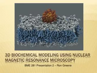
3D biochemical Modeling Using Nuclear Magnetic Resonance Microscopy BME 281 Presentation 2 – Ron Greene
The importance of visualization “The few wonders of the world only exist while there are those with the sight to see them.” – Charles de Lint (Celtic folk musician )
1978 - Kurth Würthrich used NMR to determine protein structures 1981 - Don Wiley determined the structure of the hemagglutinin protein from the surface of the influenza virus 1998 - Rod MacKinnon publishes first high-resolution structure of an ion channel 2007 - Brian Kobilka solves first structure of a human G protein coupled receptor A Brief History of 3D Molecular Modeling 1609 - Galileo Galilei develops compound microscope 1951 - Erwin Müller invents field ion microscope; sees first atoms 1964 - Aaron Klug shows Xray diffraction could be used to develop crystallographic electron microscopy 1971 - Protein Data Bank established as a repository for 3D structural data of proteins and nucleic acids
4pi – laser scanning flourescence microscope ~ 100nm scale Xray crystallography – destroys subject material, no dynamic information Nuclear Magnetic Resonance microscopy –capable of 1-2 Å resolution. Capable of imaging dynamic processes in vivo. Techniques for Molecular Imaging Crystallized proteins 4π – FGP stained live yeast H1receptor – Xray and NMR
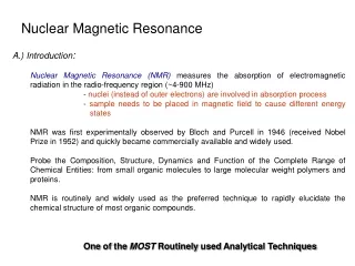
Spin – fundamental physical property of protons, electrons and neutrons Nuclear Magnetic Resonance – a phenomenon which occurs in certain atoms when exposed to magnetic field Elements with enough net spin resonate with detectable magnitudes What is nuclear magnetic resonance?
How does One Make a Nucleus resonate? When placed in a magnetic field of strength B, a particle with a net spin can absorb a photon, of frequency ω. Chemical shift affects the Larmor frequency at which a given atom resonates Chemical shift is a precise metric of the chemical environment of an atom Chemical shift After hitting with pulse to elevate energy states, NMR is measured by induction as nuclei relax to lower state Larmor frequency Gyroscopic ratio
How to react when your nmr is sending Mixed Signals NMR signal contains many components, each with its own Larmor frequency This signal, called Free Induction Delay (FID) decays with time Fourier analysis is used to transform the signal from time domain to frequency domain.
“Here’s the plan: We put the band back together, do some gigs, earn some bread, bang!” – Jake Blues
To infinity and beyond The Future: The Protein Data Bank continues to expand, including more proteins in more of their naturally occurring conformations NMR microscopy and clinical MRI merge to offer real time imaging of biological processes on an atomic scale
Magnet therapy works, man References [1] URI BME 281 BME Seminar II <www.ele.uri.edu/courses/bme281>. [2] J Puerta-Fonolla, T Vasquez-Osorio, J ruiz-Cabello, J Murillo-Gonzalez, A Pena-Melian. "Margentic resonance microscopy versus light microscopy in embryology teaching." Clinical Anatomy (2004): 429-435. [3] Oleg Yu Federoff, Miguel Salazar, Haiyong Han, ViolettaChemeris, Sean Kerwin, Laurence Hurley. "NMR-Based Model of a Telomerase-Inhibiting Compound Bound to G-quadruplex DNA." Biochemistry (1998): 12367-12374. [4] Seui Ogawa, david Tank, Ravi Menon, JuittaEllerman, Seong-Gi Kim, HellmutMerkle, KamilUgurbil. "Intrinsic signal changes accompanying sensory stiimulation: Functional brain mapping with magnetic resonabce imaging." Proceedings of the National Academy of Science (1992): 5951-5955. [5] wiki. "Nuclear magnetic Resonance." 22 October 2011. Wikipedia. 5 November 2011 <http://en.wikipedia.org/wiki/Nuclear_magnetic_resonance#NMR_spectroscopy>.