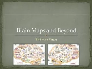
Brain Maps and Beyond
Brain Maps and Beyond. By Steven Vargas. History of Brain Mapping. Since the study of Phrenology, a now pseudoscience, people started to attribute certain brain areas with certain functions and behaviors. Phineas Gage and his frontal lobe damage, which caused him major personality changes.

Brain Maps and Beyond
E N D
Presentation Transcript
Brain Maps and Beyond By Steven Vargas
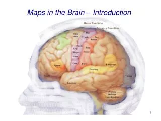
History of Brain Mapping • Since the study of Phrenology, a now pseudoscience, people started to attribute certain brain areas with certain functions and behaviors. • Phineas Gage and his frontal lobe damage, which caused him major personality changes. • Wilder Penfield and his maps for sensory and motor corticies. • These and more findings have lead to popular theories of modularity .
Broca’s Aphasia • Discovered by French neurologist Paul Broca by looking at the brains of his patients after they died. • Located in left posterior inferior frontal gyrus or Broca’s area. • It is also know as expressive aphasia. • It is categorized by deficits in language production both spoken and written • Language comprehension is roughly intact.
Patient with Broca’s Aphasia • http://www.youtube.com/watch?v=gocIUW3E-go
Wernicke’s Aphasia • Discovered by Carl Wernicke around the same time Broca found Broca’s area. • Corresponds to damage to posterior part of the superior temporal lobe known as Wernicke’s area. • Categorized by fluent unintelligible speech and language comprehension deficits. • Also known as Receptive aphasia.
Lesion correlates of conversational speech production deficits • Goal was to tap into different aspects of speech production and distinguish potentially distinct neural mechanisms. • Prompted by fact that Brocas’s area was not as predictive of symptoms of expressive aphasia in past lesion studies. By Arielle Borovsky, Ayse Pinar Saygin, Elizabeth Bates, Nina Dronkers
Methods : Subjects • Used 50 aphasiacs with different types of a aphasias including Broca’s ,Wernicke’s, and anomic aphasia. • They had unilateral lesions caused by a cerebrovascular accident. • Recruited from the San Diego community and were paid fro participating in the study.
Methods: Variables • They interviewed the subjects asking them a list of interview questions which were recorded for fluent, complex and lexical speech. • “Tokens”: the overall number of words spoken which represents overall speech fluency. I.E. “Tall – parents – often – have – tall – kids” (tokens = 6, number of words: 6) • Mean length of utterance in morphemes(MLU):used to measure grammatical complexity. I.E. “Tall – parent – s(Plu) – often – have – tall – kid – s(Plu)” (MLU = 8, number of morphemes: 8 number of utterances: 1) • Type/Token Ratio(TTR): Number of different kinds of words spoken divided by overall number words spoken to measure lexical diversity. “Tall – parents – often – have – kids (TTR = 0.83, number of unique words: 5, number of words: 6)
Methods: Variables Cont. • They created digital reconstructions of their lesions using Voxel based lesion Symptom Mapping (VLSM). • VLSM: Technique in which at each voxel, patients are divided into two groups according to whether they did or did not have a lesion affecting that voxel. • These produce colored VLSM maps that represent voxels where patients with lesions show a significantly different production score than those whose lesions are spared that voxel.
Results • Speech fluency correlated with insula , motor cortex, and superior longitudinal fasciculus(SLF). • Complexity of speech correlated with anterior portion of insula, and motor cortex. • Lexical diversity corelated with anterior medial temporal gyrus(MTG)and superior temporal gyrus (STG).
Conclusions • They found that speech fluency and complexity overlap in lesion regions. • Deficits in production of fluent and complex speech were found in lesions in motor, somatosensory cortex, anterior insula and parts of SLF as well as IFG. • Posterior MTG, STG, Angular gyrus were involved in deficits in production of semantic variety in speech. • Changes the belief that these areas were only involved in comprehension of speech.
References Images • hiddentalents.org • images.wikia.com/psychology/images/0/0a/Sensory_and_motor_homunculi.jpg • http://www.nature.com/nrn/journal/v5/n10/fig_tab/nrn1521_F1.html • http://www.profelis.org/webpages-cn/lectures/neuroanatomy_1ns.html • http://4.bp.blogspot.com/_0N6mlRJGP78/R3MQ9HpuZcI/AAAAAAAAANw/xjCgh2d7FEc/s1600-h/Gray726.png • http://knakmos.deviantart.com/art/Perception-23682233 Facts • Wikipedia Article • Arielle Borovsky, Ayse Pinar Saygin, Elizabeth Bates, Nina Dronkers, 2007,Lesion correlates of conversational speech production deficits,Neuropsychologia volume 45, issue 11.