Data Handler Display
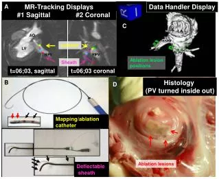
This document covers the innovative tracking, display, and mapping techniques in cardiac ablation procedures. It focuses on real-time visualization of lesion positions in various planes (sagittal and coronal) using advanced catheter technologies. Key components include the analysis of left ventricle (LV), left atrium (LA), and pulmonary veins (RIPV, RSPV) during ablation. It also touches on the importance of flexible sheaths and histological insights from pulmonary vein samples, enhancing the understanding of effective treatment strategies in electrophysiology. ###

Data Handler Display
E N D
Presentation Transcript
A MR-Tracking Displays #1 Sagittal #2 Coronal Data Handler Display C AO catheter LV LA RIPV RIPV RSPV Ablation lesion positions Sheath t=06;03, sagittal t=06;03 coronal RLPV B Histology (PV turned inside out) D Mapping/ablation catheter Ablation lesions Deflectable sheath