Action Potentials and Synapses
Action Potentials and Synapses. Action Potentials. Most nerves exhibit Action Potentials (APs) Spikes of electrical activity Almost always less than 1000 per second Almost all neurons create APs (some retinal cells only have graded potentials). Potential.

Action Potentials and Synapses
E N D
Presentation Transcript
Action Potentials • Most nerves exhibit Action Potentials (APs) • Spikes of electrical activity • Almost always less than 1000 per second • Almost all neurons create APs (some retinal cells only have graded potentials)
Potential • A potential is the potential to do work. • Work requires energy. • The Second Law of Thermodynamics says that all matter spontaneously moves towards lower energy states. That is, from regions of higher potential to regions of lower potential.
Potential • There are two major types of energy: • Potential Energy which derives from the four basic forces: • Gravity • Weak force • Strong force • Electromagnetic force • Kinetic energy from moving matter • The energy that made it move can be extracted.
Potential • The two main potential energies of interest to neurologists are: • Electromagnetic potential • Opposite charges attract, like charges repel. • Concentration potential • Flow is in direction of higher concentration towards lower concentrations until equalization.
Potential • Electromotive potential • Proportional to amount of separated charges +1.5 V Test Ref (0) -1.5 V Ref (0) Test + - + -
Potential • Potential difference is the difference in the total potentials between the two measured points. • Potential is measure in volts (V) • Think of a battery, typically 1.5 or 9 V. • A 9 V battery has a higher potential than a 1.5 V battery. • Potential is additive: 3 V 0 V
Potential • Concentration gradient • Any time there is a concentration gradient, there is force trying to move ions to area of lesser concentration. Fresh water (low NaCl) Seawater (high NaCl)
Potential • If you get nothing else out of this lecture: • Opposites attract • Concentrations equalize
Cellular Potential • Because neural cell walls are porous and there are differences in ion concentrations inside and out, the inside of a neuron has a potential with respect to the extracellular fluid. • Cellular potentials are always given with respect to the extracellular space.
Cellular Potential • Potential can be calculated by the Goldman equation, which takes into account the concentrations of the ions Na+, K+ and Cl- and their membrane permeabilities.
Cellular Potential • Normal nerve potential is about –70 mV with respect to the extracellular space.~1/20 of a battery
Nerve Cell Environment A whole lots of Na+ (sodium) 0 mV A little Cl-(chlorine) Cell Lots of K+ (potassium) -70 mV Na+ K+
Nerve Cell Membrane • Populated with ion channels • 4-7 transmembrane domain proteins • Typically only pass one type of ion • Activated by ligands, voltage, or time • Types • Passive, ionic movement by gradient • Facilitated transport • Active transport, energy powered pump
Transmembrane Transport • Simple diffusion (spontaneous) • Ions follow gradients without help • Facilitated diffusion (spontaneous) • Helper proteins assist the move • Active transport (energy required) • Normally symport – 2 ions transported at once • http://pb010.anes.ucla.edu/membrane.htm
Transmembrane Transport • Simple diffusion • Ions follow gradients without help (Na+, K+)
Transmembrane Transport • Facilitated diffusion • Larger molecules follow gradients, but have to be passively assisted to pass thru membrane. Ex: glucose transporter
Transmembrane Transport • Active transport • Molecules move against a gradient, so an energy-consuming “pump” is required. • Symport – 2 ions are moved at once. (Na+-K+ pump)
Action Potential • There are many types of “holes” or channels in the neural membrane, but the most important are: • Key-activated sodium (Na+) channels • Voltage-activated sodium (Na+) channels • Voltage-activated potassium (K+) channels • Key-activated chlorine (Cl-) channels • An ATP-powered sodium-potassium pump (symport)
Action Potential • Cell at –70 mV. Normally all channels are closed. • “Something” opens keyed Na+ channels. • Na+ enters the cell, depolarizing it. • When potential reaches threshold (about -40 mV) at the axon hillock, many more voltage-controlled Na+ channels open causing a flood of Na+ depolarizing the cell to about +30 mV. • Na+ channels close and voltage-controlled K+ channels open, K+ flows out repolarizing the cell. • Na+/K+ pump restores equilibrium.
Action Potential • AP propagation • The local depolarization triggers the voltage controlled pores in the adjacent region, causing another depolarization. • The depolarization progresses along the length of the axon.
Action Potential • At the terminals, the depolarization allows Ca++ to enter the terminal and trigger fusion of neurotransmitter vesicles to the membrane. • Neurotransmitter is released via exocytosis.
Action Potential • Soluble NSF Attachment protein REceptors (SNAREs) keep vesicles in close proximity to membrane. • Ca++ causes SNAREs to complete fusion.
Action Potential • Amino acid and amine neurotransmitters are synthesized in the terminal button. • All others, including peptides and larger proteins, are manufactured in the cell body and transported to the terminal button at speeds of up to 1 meter per day. • The latter may suffer periods of insufficient neurotransmitter.
Action Potential • “All or Nothing” firing • Once threshold is reached, the nerve will always fire
Action Potential • Refractory periods • Absolute • About 1 mSec, cannot fire again. • Due to time-activated gate in Na+ channels. • Relative • About 5-10 mSec, can fire again with more stimulation. • Due to undershoot.
Action Potential • Firing rate limited to about 1000 Hz • Due to absolute refractory period • Firing rate varies with stimulation, but almost never ceases entirely • Non-quiet presynaptic neurons • Random molecular motion • Mechanical forces • Ionic leakage through nerve wall
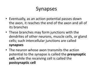
Synapse • The space between the terminal button of a presynaptic nerve and a dendrite, dendritic spine, soma or axon of the postsynaptic cell.
Synaptic Transmission • The neurotransmitter diffuses across the synapse and binds to receptors on the postsynaptic nerve dendrites. • When enough stimulation has been received, the postsynaptic nerve fires an AP. • Presynaptic autoreceptors limit neurotransmitter release. • Excess neurotransmitter is destroyed or reuptaken into the presynaptic nerve.
Synaptic Transmission • “Something” opens receptor Na+ channels • Neurotransmitters • From presynaptic cell • Very localized action • Neuromodulators • From nearby cells • Local area, longer term action • Hormones • Transported via blood, not neurons • Systemic, long term action
Synaptic Transmission • Inhibition • Similar to how opening Na+ channels allows the nerve to depolarize towards threshold, inhibitory channels allow negative ions to enter and hyperpolarize the cell, moving potential farther away from the threshold. • Usually opens Cl- channels • GABA, Serotonin (5-HT), etc.
Synaptic Transmission • Excitory Post- Inhibitory Post-Synaptic Potential Synaptic Potential EPSP IPSP
Synaptic Transmission • Excitatory and Inhibitory Inputs • Sum spatially and temporally. • Many thousands of inputs usually necessary to reach threshold.
Action Potential • Conduction Velocity • The larger, the faster! • < 1 m/s for small, unmyelinated neurons • > 50 m/s in large myelinated neurons
Myelin • Conduction can be speeded by myelin • Segmented non-conductive sheath surrounding the neuron. • No (few) ion channels underneath the sheath. • High concentration of ion channels in the nodes between sheath segments. • AP “jumps” from one node to the next, bypassing propagation delays (known as saltatory conduction). • Can speed conduction by 100x.
Myelin • Schwann Cells in PNS • Tightly wrapped around axon. • Each segment about 1 mm, no ion channels. • Nodes of Ranvier in between.
Myelin • Oligodendrocytes in CNS • Each cell wraps several nerves. • More compact than individual Schwann cells. • Also segmented with nodes.
Postsynaptic Effects • Three main types of effects: • Direct effect on ionic channels • Second messenger systems • Direct gene activation
Postsynaptic Effects • Direct effect on ionic channels • Neurotransmitter binds to receptor channel causing a conformational change which opens the channel to allow the ion to pass. • Quickest acting and shortest acting effect. • Neurotransmitter remains in synapse and is metabolized or reuptaken.
Postsynaptic Effects • Second messenger systems • A guanine sensitive protein (G-protein) is linked to the receptor. • Neurotransmitter binds to receptor channel causing G-protein to activate a catalytic enzyme in the membrane. • The enzyme produces a “second messenger.” • The 2nd messenger can affect other receptors or can directly influence protein synthesis. • Effects are slower, but longer lasting.
Second Messengers • The most common 2nd messengers are: • Cyclic Adenosine Monophosphate (cAMP) • Inositol Triphosphate (IP3) • Diacylglyceride • 2nd Messengers: • AMPLIFICATION • PROLONGED EFFECT
Direct Gene Activation • Primarily lipid-soluable steroids • Pass through the cell and nuclear membranes. • Led to DNA by chaperonin-receptor complexes. • Receptor binds to acceptors on chromatin. • Transcription of specified protein begins. • mRNA goes to rough ER for protein synthesis.
Neurotransmitters • Trigger postsynaptic nerves • Major classes of neurotransmitters • Amino Acids • Monoamines • Acetylcholine • Soluable Gasses • Peptides • Steroids