Nerve Signals : Action Potentials
340 likes | 609 Vues
Nerve Signals : Action Potentials. Resting Potential. ‘Resting’ refers to the neuron not conducting a signal (not conducting an action potential ).

Nerve Signals : Action Potentials
E N D
Presentation Transcript
Resting Potential ‘Resting’ refers to the neuron not conducting a signal (not conducting an action potential). Inactive (non-signaling) neurons maintain an electrical potential (a relative voltage difference) across their axomembranes of ~ -65 mV. Axomembranes are said to be polarized. By convention, the ECF voltage is called zero. • This indicates that, relatively speaking, the inside of the axon is more negative (or less positive) in charge than the outside of the axon (ECF). • Such a potential is measured by an oscilloscope, which is a fancy voltmeter designed to test neurons. • The difference in charge (electrical potential or axomb polarization) is attributable to a number of factors:
In no particular order of importance: i. A Na+/K+ pump (carrier protein) maintains an unequal distribution of Na+ and K+ across an axomembrane – there exists a higher [Na+] outside the neuron (in the ECF), and a higher [K+] inside the neuron (in the axoplasm). This, in itself, does not contribute to the negative cell potential, but the fact that the pump actively moves 3 Na+ out for every 2 K+ in (this is done primarily after nerve signals to reset the neuron) contributes to there being, at any one time, more cations outside than inside the neuron; thus contributing to the relative negative polarity across the axomembrane. The pump, which moves these ions against their concentration gradients, serves to maintain the steep concentration gradients mentioned with respect to Na+ and K+ (important for nerve signals).
ii. Na+ and K+ possess specific channel proteins (more for K+) that allow for a slow leakage of ions down their concentration gradients. K+ leaks out about 50x faster than Na+ leaks in promotion of negative resting potential (there are very few of these ‘open’ channels compared to the soon-to-be-mentioned ‘gates’ that open during an action potential). The pump acts to continuously correct leakage. iii. There exist plenty of large anions within the neuron (proteins, amino acids, sulfate, phosphate), which cannot move out of the cell (either too large or possessing no specific channel proteins), thus creating a perpetual contribution of negative charge inside. iv. The [Cl-] is greater outside than inside (which might have you wondering…), but at least there is some (albeit much less than outside) Cl- inside; this, in contrast with the large anions mentioned in (iii), where they only exist inside the cell. See fig. 17.4(a) p. 325.
Methods of Ion Transfer Across Axomembrane • Channel Protein Diffusion: this refers to the leakage of Na+ and K+ ions; • Active Transport – Na+/K+ Pump: moves ions against their conc. gradients to ‘reset’ neuron for another action potential (ie. re-creates steep gradients mentioned earlier); • Protein Gates (Diffusion): specific, one-way proteins (gates) – two types: Na+ gates (open to allow Na+ into axon (down its conc. gradient)), K+ gates (open to allow K+ out of axon (down its conc. gradient)) – these will be discussed in detail during analysis of action potentials; however, it is important to point out that these gates, when opened, serve as the primary mechanism by which Na+ and K+ move into/out of the neuron as they vastly outnumber the ‘always open’ channels mentioned earlier. An action potential requires a flood of ions in either direction – thus the need for the numerous gates.
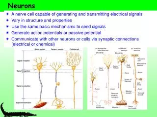
Action Potential (the SIGNAL!) • The signal transmitted along the length of a neuron, from a dendrite, cell body, or axon hillock to the synaptic endings of an axon, is an electrical signal that depends on a rapid, localized flow of ions across the neuron’s mb. • This signal (flow of ions across the mb) then travels in a perpendicular direction along a neuron (‘domino’ effect). • All cells possess a membrane potential of some kind, but only neurons (and muscle cells) have the ability to generate changes in that potential (and thus, generate a signal).
A stimulus is received by a dendrite (or a cell body); the stimulus can be pressure, temperature, light (eyes), or chemical and it is received by specific receptors on the dendrites. • Chemical stimuli usually come from other neurons, whereas the other stimuli arise from the environment. • The stimulus is passed to the cell body, which then integrates it. If the stimulus is ‘weak’, it will not reach the axon hillock; if it is ‘medium’-‘strong’, it is passed on to the axon hillock. • If the stimulus is strong enough, it will cause the axomembrane of the axon hillock to depolarize to a certain degree (ie. the axoplasm will become less negative in charge). If that certain degree of depolarization reaches a certain threshold, an action potential (AP) will result. • How does the depolarization occur? See fig. 17.4(b) p.325
Steps: 1. Resting Potential – as described earlier… 2. DEPOLARIZATION -- stimulus reaches axon hillock and causes Na+ gates to open. A stronger stimulus initially opens more gates, and vice versa. This allows Na+ to move into the axoplasm (across the axomembrane) down its concentration gradient (and due to the fact that the axoplasm is negative in charge compared to Na+’s charge – opposites attract!). If the stimulus is strong enough, threshold potential will be reached (-55mV to –40mV, depending on text; we’ll go with our text (-40mV)) and ALL remaining Na+ gates open (example of positive feedback) so that Na+floods the axoplasm. Na+ equilibrium (with respect to charge and concentration) is reached when the potential across the axomembrane (axoplasm charge) equals +40mV. Depolarization (APs) are referred to as ‘All-or-None’ meaning that threshold is either reached, or not reached.
So, if the stimulus is not strong enough to open a sufficient amount of Na+ gates, threshold will NOT be attained, and the signal ‘dies’. 3. REPOLARIZATION – once Na+ equilibrium is attained, the Na+ gates close. This signals K+ gates to open and K+ floods OUT of the axoplasm into the ECF (due to conc. gradient and electrical gradient). K+ equilibrium is attained at ~ -80-85mV (called the ‘undershoot’). The K+ gates then close. 4. REFRACTORY PERIOD – consists of two processes: a. excess K+ in ECF diffuses away to re-establish -65mV resting potential.
b. Na+ gates are unable to open for a certain period of time (this disallows a backward nerve signal whenever it is initiated at the axon hillock). *once depolarization is complete, and the refractory period passes, that particular section of the axon is able to ‘fire’ again – this is due to the fact that, even though ions changed places and did not return, only a small percentage of the TOTAL Na+ and K+ swapped locations. A section of an axon may fire multiple times before it requires a Recovery Period (Ion Redistribution period), where both gates shut down so that the Na+/K+ pump can get the ions back to where they originally came from.
All-or-None Depolarization • Action potentials are all of equal strength (ie. if a stimulus is received, it either reaches threshold, and therefore +40mV (Na+ equilbrium) or it does not and resting potential is maintained). • So, how does a stronger stimulus affect nerve firing? • A stronger stimulus simply produces more APs per unit time than does a weaker stimulus (that attains threshold). • Thus, the refractory period for a neuron conducting a stronger signal (more APs) is shorter than that of a neuron conducting a weaker signal (less APs). • This is important during stressful situations as, eventually, neurons need a recovery period (pump working feverishly), yet the signal must persist – this is where hormones come into play to maintain a signal.
Propagation of an Action Potential (Domino Effect) • An AP (depolarization/repolarization) is a localized event that tends to originate at the axon hillock. • APs need to ‘travel’ along an axon to carry the signal – the ‘traveling’ is actually due to simple diffusion of Na+ within the axoplasm. • Once Na+ floods the axon (depolarization), the ions diffuse in either direction in the axon (from higher to lower conc.). • Na+ that diffuses forward in the axon allows the axoplasm in this ‘non-stimulated region’ to reach threshold (cations raise voltage to –40mV), which triggers the Na+ gates in this ‘new’ axon region to open, so that more Na+ may flood across the axomembrane. • The Na+ that flows backward has no effect since that region of the axon is experiencing the refractory period where Na+ gates can not open for a period of time – thus, APs cannot travel backward.
Myelin • Myelin is a lipid that serves as a neuron insulator by wrapping around axons (and dendrites in sensory neurons). • It is composed of Schwann Cells (aka oligodendrocytes or neurolemmocytes). • Myelin serves to increase the speed of an AP’s (nerve impulse’s) propagation by simply acting as a physical barrier to ion flow (both Na+ and K+). • Axons are not continuously covered by myelin; the myelin sheath is interrupted at regular intervals by nodes (‘naked’ or ‘bare’ axon regions) called Nodes of Ranvier. • The specific Na+ and K+ gates congregate at the nodes in myelinated neurons – therefore, depolarization and repolarization may only occur at the nodes. • Thus, APs propagate (‘jump’) from node to node (known as Saltatory Conduction). • Non-myelinated neurons’ APs travel much more slowly…
Communication between Neurons and OtherCells • When an AP arrives at the many synaptic endings (axon bulbs), the ‘signal’ must somehow be passed on to another cell for its desired effect to occur (remember: synaptic endings DO NOT touch target cells). • There are three possible target cells: • Another Neuron – the connection is called a synapse; • Muscle Cell – the connection is called a neuromuscular junction (synapse); • Gland or Organ Cell – the connection is called a neuroglandular junction (synapse).
Major focus will be on the synapses between neurons, which conduct signals from an axon’s synaptic endings to the dendrites (or cell bodies, in some cases) of the next neuron in a signal pathway. • The transmitting cell (the cell trying to ‘pass on’ the AP) is called the presynaptic cell and it possesses its own membrane, the presynaptic membrane. • The receiving cell (the cell destined to ‘receive’ the AP) is called the postsynaptic cell and it possesses its own membrane, the postsynaptic membrane. • The space between the two neurons is known as the synaptic cleft (really, it’s just ECF with a max. width of 0.2 micrometers).
There exist two types of synapses: Electrical and Chemical we will focus primarily on the chem. • Electrical Synapses: allows APs to spread directly from pre- to postsynaptic cells. The cells are connected by gap junctions (recall SA node signaling in heart for atrial contraction), which allow for direct ion transfer between neurons (much faster, but more energy/coordination to control); chemical synapses FAR outnumber electrical synapses in the body.
Chemical Synapses (aka ‘Synapses’) see fig. 17.5 p. 326 • The synaptic endings of presynaptic cells contain vesicles that are full of tiny chemical ‘messengers’ known as neurotransmitters; each vesicle is attached to the presynaptic membrane by a special, thread-like contractile protein (fiber) (primarily composed of actin – a microfilament). • The postsynaptic membranes possess multiple, specific protein receptors for these neurotrans-mitters to bind to; a different type of neurotrans-mitter will bind to a different receptor and will produce a different resulting signal.
The Transmission Process: • When an AP arrives at a synaptic ending, Na+ floods into the axon bulb, which serves to signal different channels to open (Ca2+ channels), which are only located in the synaptic endings. Ca2+, which predominantly exist within the synaptic cleft, then flood the bulb (of course, K+ leaves to repolarize). • Ca2+ bind to the contractile proteins and cause them to CONTRACT and shorten, thus pulling the vesicles to the presynaptic membrane. • Exocytosis occurs at the mb to release the neurotransmitter into the synaptic cleft (many mitochondria exist within the axon bulb in order to promote this active process). Once enough vesicles have released their neurotransmitters, the ATP from the mitochondria helps to actively transport the Ca2+ ions back out of the bulb into the synaptic cleft.
4. Chemical neurotransmitters simply diffuse across the synaptic cleft into the vicinity of the postsynaptic membrane (cell). 5. The neurotransmitters bind with their specific, respective protein receptors that are immersed within the postsynaptic membrane. 6. One of six outcomes may occur depending on the type of postsynaptic cell and the type of neurotransmitter/receptor combo at work. Recall the three types of postsynaptic cells: another neuron, a muscle cell, or a gland/organ cell. The two different neurotransmitters that we talk about are norepinephrine (excitatory) (aka ‘noradrenalin’) and acetylcholine (inhibitory)…thus, they promote contrasting effects upon their target postsynaptic cells:
Norepinephrine initiates an excitatory postsynaptic potential (EPSP). Acetylcholine initiates an inhibitory postsynaptic potential (IPSP). 7. Receptors release the neurotransmitters back into the synaptic cleft after their signaling function is completed. To prevent perpetual stimulation or inhibition, these ‘used’ neurotransmitters need to be removed from the cleft. The following outlines how this is done: • They are reabsorbed (endocytosis) by the presynaptic cell’s synaptic ending (axon bulb). • They simply diffuse away from the receptors.
iii. They are destroyed by enzymes that exist on the postsynaptic membrane (the enzymes are activated when the neurotransmitter is released back into the cleft): Monoamine Oxidase destroys norepinephrine; Acetylcholinesterase destroys acetylcholine. * Some drugs work to promote a continued existence of neurotransmitters within the cleft so that their excitatory/inhibitory effects occur over a lengthy period of time (eg. Cocaine = excitatory; Heroin = inhibitory).
Summation/Integration at Synapses • A single neuron possesses numerous dendrites along with the cell body, each of which synapse with thousands of synaptic endings of other neurons in a relatively small volume of space (if you recall, many neurons are bundled into nerves). • Because the cleft is a ‘sea’, of sorts, many different neurotransmitters might be ‘floating’ around affecting many synapses. • Thus, a postsynaptic cell might be receiving a differing combination of excitatory and inhibitory neurotransmitters (eg. all of one type, or a mix of each), which possess the ability to cause either depolarization (opening of Na+ gates) or hyperpolarization (opening of K+ gates).
The cell body and axon hillock then sum up (integrate) the ions received (Na+) and lost (K+) to ‘decide’ if threshold is reached (see fig. 17.6 p. 327). • If threshold is not reached, no AP is generated in the axon hillock; • If threshold is reached, an AP is generated and continues at the same strength along the length of the axon to the postsynaptic cell’s synaptic endings. • Two types of summation: • Temporal Summation: neurotransmitter of one type released rapidly from one presynaptic nerve (bundle of neurons) so as to trigger a particular summed response; • Spatial Summation: several different synaptic endings belonging to different presynaptic cells (nerves) stimulate a postsynaptic cell at the same time and have an additive effect.