Download
1 / 142
1.53k likes | 2.8k Vues
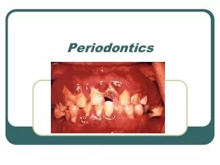
Pre-clinical Periodontics. Dr Jamal Naim PhD in Orthodontics. Introduction. Definitions. Carranza's - clinical periodontology. Periodontal Disease. Periodontology. It is a three quarter course, one preclinical and two clinical courses.

E N D
Pre-clinical Periodontics Dr Jamal Naim PhD in Orthodontics Introduction
Definitions Carranza's - clinical periodontology
Periodontology • It is a three quarter course, one preclinical and two clinical courses. • The Completion of preclinical course should enable undergraduates to: • Screen for risk of periodontal diseases. • Apply charting systems for management and monitoring patients at risk of periodontal disease. • Carry out simple treatment procedures required for stabilizing periodontal disease.
Preclinical Periodontology • Element Objectives: • A student should acquire basic information and understanding of: • The Normal Periodontium • Classification & Epidemiology • Classification of diseases of the periodontium and epidemiology of gingival & periodontal disease • Etiology of Periodontal diseases • Gingival inflammation & its clinical features, • Treatment of Periodontal disease
Preclinical Periodontology • Clinical, Radiographic & advanced diagnostic techniques, • Understand the different processes healing, repair and tissue loss occurring as a result of gingivitis and periodontal diseases. • Learn basic instrumentation techniques for supra and subgingival debridement. • On completion this course the students should have worked on natural extracted teeth embedded in casted models to demonstrate their ability for scaling of supra-gingival calculus and polishing of crown and scaled roots.
Introduction • The oral mucosa is located between skin and gastrointestinal mucosa and shows some of the properties of each. • Like skin the oral mucosa consists of two tissue components: • A covering epithelium and • underlying connective tissue
Introduction • The main functions of the oral mucosa are: • Protection • Sensation • Secretion • Thermal regulation
Introduction • There are three main types of the oral mucosa, identified according their primary function: • Masticatory mucosa • Lining mucosa • Specialized mucosa • The larger part of the OM is lining mucosa (60%), followed by the masticatory (25%) and specialized mucosa (15%)
Oral Mucosa MASTICATORY SPECIALIZED DORSUM OF TONGUE GINGIVA, HARD PALATE LINING LOOSELY ATTACHED FIRMLY ATTACHED LIPS , CHEEKS, VENTRAL SURFACE OF TONGUE SOFT PALATE ALVEOLAR MUCOSA, FLOOR OF MOUTH VESTIBULAR FORNIX
Epithelial maturation • Cells that are driven from progenitor cells and ready for maturation passes the entire epithelium to form the protective layer. • In general maturation in the oral cavity follows two main patterns: • Keratinization • Nonkeratinization
Epithelial maturation The turnover time is the period of time is needed by a cell to divide and pass through the entire epithelium. The turnover time is 52 to 75 days in skin, 41 to 57 days in the gingiva and 25 days in the cheek.
Keratinized epithelium Keratinization or cornification is the formation of a surface of keratin. Such process is seen in the oral mucosa of the palate, gingiva and in some regions of the tongue dorsum. A keratinized epithelium shows in histological sections a number of layers (strata).
Keratinized epithelium • The basal layer - stratum basale – • Theprickle cell layer- stratum spinosum – • The granular layer - stratum granulosum – • The keratinized layer - stratum corneum –
Non-keratinized epithelium Non-keratinization occurs in regions with less mechanical influences to the OM, such as cheek, lips, underside of the tongue and the soft palate. Non-keratinized epithelium is usually thicker than keratinized epithelium. No sudden changes in the cells above the st. spinosum occur in non-keratinized epithelium, and the outer half of the tissue is divided into two zones:
Epithelial maturation • Intermediate layer - stratum intermedium - • Superficial layer – stratum superficiale –
Epithelial maturation In so called parakeratinized mucosa, such as parts of the hard palate and the gingiva, in the surface layer the nuclei are shrunken and retained in many or all squames. Also keratohyaline granules are present in this layer. Such phenomenon is a normal event in the oral epithelium, but not true for the epidermis, where parakeratinization is associated with diseases such as psoriasis.
Definitions Please read chapter (Periodontium) in OB Carranza's - clinical periodontology
Gingiva • Gingiva is that portion of the oral mucosa that covers the tooth-bearing part of the alveolar bone and the cervical neck of the tooth • Morphologically gingiva is divided into: • Free gingiva • Attached gingiva • Gingival sulcus • The interdental papilla.
Free gingiva • The free gingiva is a relatively mobile tissue surrounding the gingival sulcus. It covers approximately 1.0 to 1.5 mm of the tooth surface. • It extends along the cervical level of the tooth at the labial, buccal and lingual surfaces and interdental. • It is freely movable and extends from the gingival margin to the free gingival groove
Free gingiva In about one third of all individuals, a shallow freegingival groove runs parallel to the gingival margin along a line that is located roughly at the junction between the free gingiva and the attached gingiva. The free gingival groove is positioned at the height of the CEJ at the bottom of the gingival sulcus or slightly below
After completed tooth eruption, the free gingival margin is located on the enamel surface approximately 1.5 to 2 mm coronal to the cemento-enamel junction.
Interdental papilla The free gingiva that occupies the interdental spaces coronal to the alveolar crest is the interdental gingiva/papilla The shape of the interdental papilla is determined by the contact relationships between the teeth, the width of the approximal tooth surfaces, and the course of the CEJ.
Interdental papilla In anterior regions of the dentition, the interdental papilla is of pyramidal form while in the molar regions, the papillae are more flattened in buccolingual direction interdental papilla in the posterior region interdental papilla in the incisor region
Interdental papilla In the premolar/molar regions of the dentition, the teeth have approximal contact surfaces rather than contact points. In the posterior region the papilla is composed of an oral and a vestibular papilla joined by an interdental col. interdental papilla in the posterior region
Interdental papilla The col region is covered by a thin non-keratinized epithelium
Interdental papilla In cases where no contact point (e.g. diastema) the interdental papilla is reduced in height.
Attached gingiva • The bulk of the gingiva is firmly attached to the tooth and the alveolar bone by well-developed collagenous fiber bundles • It extends from the free gingival groove to the mucogingival junction which separates the attached gingiva from the alveolar mucosa. • Its surface shows stippling - "orange peel" appearance-.
Attached gingiva • The epithelium of the attached gingiva is keratinized or para-keratinized • The lamina propria contains numerous collagen bundles attaching the tissue to the periosteum • The collagen bundles cause • the stippling, and the absence • of the stippling don’t denote • always inflammation.
Attached gingiva The width of attached gingiva varies for each tooth. The attached gingiva is wider in the maxilla, especially on the labial surfaces of the incisors, and narrowest over the buccal surfaces of the mandibular canines and first premolars and the lingual surfaces of the mandibular incisors. The width of the attached gingiva varies from 1.0 to 6.0 mm. The width of the keratinized gingiva (attached gingiva plus the free gingiva) may vary from 1.0 mm to 9.0 mm
Attached gingiva There is no significant change between free gingiva and attached gingiva, only the stippling is not more present The attached gingiva depressed between the eminencies of the sockets forming grooves called interdental grooves
Attached gingiva Lingual aspect of the mandible showing the tightly adhering gingiva (G) and the adjacent non-keratinized alveolar mucosa (AM) that lines part of the alveolar process and floor of the mouth (F). MGJ, mucogingival junction.
Attached gingiva This view of the hard palate shows the absence of a muco-gingival junction on the palatal aspect. Instead, the masticatory mucosa of the gingiva (G) blends imperceptibly with the masticatory mucosa of the hard palate (PM). Note the palatal rugae (RR), the ridges behind the anterior teeth, on either side of the incisive papilla (IP).
Gingival sulcus It is a shallow groove or crevice lined by non-keratinized epithelium and its bottom present at the point of separation of the attached epithelium from the tooth. Its average depth is about 1-2mm.
Gingival sulcus In clinically healthy gingiva there is no "gingival pocket" present but the gingiva is in close contact with the enamel surface. when a periodontal probe has been inserted in the tooth/gingiva interface a "gingival crevice" artificially opens approximately to the level of the cemento-enamel junction. After completed tooth eruption, the free gingival margin is located on the enamel surface
Epithelial Components of the Gingiva • The epithelium covering the free gingiva is differentiated as follows: • oral epithelium (OE), which faces the oral cavity • oral sulcular epithelium (OSE), which faces the tooth without being in adhesion with the tooth surface • junctional epithelium (JE), which provides the contact between the gingiva and the tooth
Epithelial Components of the Gingiva The junctional epithelium forms the dento-epithelial junction apical to the sulcus. Its coronal end forms the bottom of the gingival sulcus and is overlapped by the sulcular epithelium.
Epithelial Components of the Gingiva 3. Junctional epithelium: It is the stratified non-keratinizing epithelium, that surrounds the tooth like a collar with a cross-section resembling a thin wedge. It is attached by one broad surface to the tooth and by the other to the gingival connective tissue. Rete pegs are lacking in the junctional epithelium.
Epithelial Components of the Gingiva • These epithelia differ from one another in their function and, therefore, in some of their histological characteristics: • The size of the cells in the junctional epithelium is, relative to the tissue volume, larger than in the oral epithelium. • The intercellular space in the junctional epithelium is, relative to the tissue volume, comparatively wider than in the oral epithelium. • The number of desmosomes is smaller in the junctional epithelium than in the oral epithelium.
Epithelial Components of the Gingiva • The oral epithelium is the stratified, squamous keratinized • The sulcular epithelium is the stratified, squamous epithelium, non-keratinized or parakeratinized • A characteristic morphologic feature of the oral epithelium and the oral sulcular epithelium is the presence of the epithelial ridges -rete pegs-, while these structures are lacking in the junctional epithelium.
Epithelial Components of the Gingiva The junctional epithelium has 2 basal laminas, one that faces the tooth (internal basal lamina) and one that faces the connective tissue (external basal lamina). The proliferative cell layer responsible for most cell divisions is located in contact with the connective tissue, i.e. next to the external basal lamina.
Epithelial Components of the Gingiva The junctional epithelium is more permeable than the oral or sulcular epithelium. It serves as the preferential route for the passage of bacterial products from the sulcus into the connective tissue and for fluid and cells from the connective tissue into the sulcus. Arrows indicate path taken by cells and fluids between the sulcus and the gingival connective tissue
Epithelial Components of the Gingiva The term epithelial attachment: refers to the attachment apparatus, i.e. the internal basal lamina and hemidesmosomes, that connects the junctional epithelium to the tooth surface. This term is not synonymous with junctional epithelium which refers to the entire epithelium.