Download
1 / 35
370 likes | 893 Vues
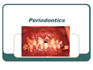
Pre-clinical Periodontics. Dr Jamal Naim PhD in Orthodontics. Etiology of periodontal diseases II. Dental biofilm. Microbial biofilms is composed of: Bacteria A matrix. Bacteria in dental biofilm. Types of Bacteria found will vary depending on: Age: immature vs. mature

E N D
Pre-clinical Periodontics Dr Jamal Naim PhD in Orthodontics Etiology of periodontal diseases II
Dental biofilm Microbial biofilms is composed of: • Bacteria • A matrix
Bacteria in dental biofilm Types of Bacteria found will vary depending on: • Age:immature vs. mature • Site:supragingival vs subgingival; (smooth surface/pit & fissures/interproximal) • Presence of disease: cariesvs. gingivitis vs. periodontal disease
Bacteria in dental biofilm Types of Bacteria found will vary depending on: • Nutrient supply: eg: Availability of nutrients from • Saliva • Gingival crevicular fluid • Dietary intake of host - fermentable sucrose • Oxygen & pH levels
Plaque matrix of dental biofilm Plaque matrix of dental biofilm consists of: Mainly - bacterial products (bacteria, dead bacteria, bacterial products = EPS, toxins, acids) & Some - host material (salivary glycoproteins, gingival fluid/exudate) - Food debris, epithelial cells, leukocytes
Plaque matrix of dental biofilm Chemical composition: • Proteins • Lipid • Carbohydrates -bacterial extrapolysaccharides (EPS) ie: glucans/fructans • Inorganic compounds – calcium, phosphate and fluoride containing
Plaque matrix of dental biofilm EPS - have an important role in the dental biofilm: • Allow bacteria to adhere and aggregrate. • Coat the bacterial cell and help protect it. • EPS give white colour to the dental plaque.
How does dental biofilm form ? Stage 1: Initial colonisation Addition of new bacteria Stage 2:Rapid bacterial growth Multiplication Stage 3: Remodelling stage/maturation Accumulation of bacterial and host products
Stage 1: Initial colonisation • First minutes to hours: covering of absolutely clean teeth by a 0.1 – 0.8 µm pellicle composed of salivary glycoproteins • Within 8 hours: gram-positive bacteria form primary colonies upon the pellicle (1-20 cells in thickness, streptococcus mutans and actinomyces species), • Non pathogenic organisms. • Aerobic (tolerate O2) bacteria.
Stage 2: Rapid bacterial growth • Multiplication occurs over the next 8 - 48 hours. • Organisms attached to the pellicle, multiply by cell division. • EPS is produced by bacteria more bacteria adhere • Plaque has doubled in mass in 2 days.
Stage 3: Remodeling/Maturation • 48 hours + • Accumulation of bacterial and host products • Maximum numbers of organisms, increasing complexity • Rapid changes occur in first 4-5 days, g- cocci, g+ and g- rods and filaments become established • plaque becomes stable around 21st day (3 weeks) with anincrease in filamentous organisms So now we speak about mature supragingival plaque
Supragingival plaque • The metabolic products stimulate a PMN migration and sulcus fluid flow in the tissue. • This the first defense mechanism of the body to wall off invading bacteria. • With the time, the JE become less resilient permitting the ingress of bacteria between tooth and JE, a gingival pocket develops.
Pristine VS normal gingiva
Supragingival plaque Development of a pocket
Characteristics of Mature plaque • more Gram negative (don’t attach initially due to poor attachment) • more anaerobic (O2 intolerant) bacteria • many of which arefilamentous bacteria • greater proportion of pathogenic microorganisms
Subgingival plaque • A dense layer of varying thickness adheres to the root surface. • Its composition resembles that of the supragingival plaque, g+ cocci, g+ filaments and actinomyces species • If mineralized, it becomes subgingival calculus. Subgingival plaque
Subgingival plaque • Freely moving bacterial accumulations (swimmers) are observed near soft tissue surface. • The swimmers contains g- anaerobes, cocci, spirochetes and rods. • They are pathogenic and increase sharply in number in acute lesions and play an important role in the progression of periodontitis.
Diseases caused by dental biofilms?? • Caries: Steptococcus mutans (& lactobaccilli) • Periodontal disease: Bacterial by-productsinitiate a host response (neutrophils react against these bacterial products).Results in Inflammation of gingival tissues. Bacterial species (usually anaerobic bacteria) presentin plaque is more important than the amount ofplaque.
Diseases caused by dental biofilms?? • Osteomyelitis • Otitis media • Etc.
Clinical detection of dentalbiofilms • Visual • Use of plaque disclosing agents: eg, erythrocin (pink-red) ; iodine solution (brown-black)
Removal of dental biofilm • Not with water or rinsing alone • Mechanically needs to be removed by toothbrushing & flossing
3. Dental calculus • Dental plaque in which mineralisation has involved both the plaque matrix and the micro-organisms. • free surface usually unmineralised plaque If the supragingival plaque become mineralized, it becomes supragingival calculus
If the supragingival plaque become mineralized, it becomesSubgingival calculus
Calculus composition • 70-90% inorganic: CaPO4, Ca(PO4)2, CaCO3 and etc. • Rest are organic: protein-polysaccharides, desquamatised oral epithelial cells, leukocytes and carbohydrates
Calculus formation • Calculus is mineralized plaque • Mineralisation begins between 1. and 14. day • saliva is supersaturated with Ca and PO4 ions ad forms the source of minerals for supragingival plaque • Gingival cervical fluid builds the source of minerals for subgingival plaque
Clinical features of dental calculus • Location : • Supra- and subgingival • near openings of salivary ducts (buccal to upper M1 and lingual lower anterior) • Colour and hardness - depends on location • Supra = white or yellow, loosely attached • Sub = black/brown, hard and firmly attached • Thickness - subgingival is thinner due to location • Removal - professionally only
SUBGINGIVAL CALCULUS BEFORE AND AFTER PROFESSIONAL SCALING
SUBGINGIVAL CALCULUS On x-ray
Factors influencing plaque retention Natural factors:
Factors influencing plaque retention Iatrogenic factors: • Subgingival filling margins • Restorative procedures • Clasp design in prostho • Subgingival crown margins
Food debris, food impaction, Materia Alba Food debris adhere lightly to teeth and mucosa and can be easily rinsed away with water. Food impaction occur in spaces when foodstuff become trapped, but can be removed mechanically. Materia alba consists of: • Bacteria • Desquamatised oral epithelial cells • Food debris • loosely bound - easily removed by water spray