Download
1 / 43
470 likes | 948 Vues
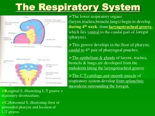
The Respiratory System – Part I. Chapter 13 BIO 160 Kelly Trainor. Objectives. Name the organs of the respiratory system and describe their function. Functions of the Respiratory System. Gas exchanges between the blood and external environment Occurs in the alveoli of the lungs

E N D
The Respiratory System – Part I Chapter 13 BIO 160 Kelly Trainor
Objectives Name the organs of the respiratory system and describe their function
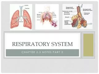
Functions of the Respiratory System • Gas exchanges between the blood and external environment • Occurs in the alveoli of the lungs • Passageways to the lungs purify, humidify, and warm the incoming air
Organs of the Respiratory System • Nose • Pharynx • Larynx • Trachea • Bronchi • Lungs • Alveoli
The Nose • Only externally visible part of the respiratory system • Air enters the nose through the external nostrils (nares) • Interior of the nose consists of a nasal cavity divided by a nasal septum
Anatomy of the Nasal Cavity • Olfactory receptors are located in the mucosa on the superior surface • The rest of the cavity is lined with respiratory mucosa that • Moisten air • Trap incoming foreign particles • Lateral walls have projections called conchae • Increase surface area • Increase air turbulence within the nasal cavity • The nasal cavity is separated from the oral cavity by the palate • Anterior hard palate (bone) • Posterior soft palate (muscle)
Pharynx (Throat) • Muscular passage from nasal cavity to larynx • Three regions of the pharynx • Nasopharynx—superior region behind nasal cavity • Oropharynx—middle region behind mouth • Laryngopharynx—inferior region attached to larynx • The oropharynx and laryngopharynx are common passageways for air and food • Tonsils of the pharynx • Pharyngeal tonsil (adenoids) are located in the nasopharynx • Palatine tonsils are located in the oropharynx • Lingual tonsils are found at the base of the tongue
Larynx (Voice Box) • Routes air and food into proper channels • Plays a role in speech • Made of eight rigid hyaline cartilages and a spoon-shaped flap of elastic cartilage (epiglottis)
Structures of the Larynx • Vocal folds (true vocal cords) • Vibrate with expelled air to create sound (speech) • Glottis—opening between vocal cords • Thyroid cartilage • Largest of the hyaline cartilages • Protrudes anteriorly (Adam’s apple) • Epiglottis • Protects the superior opening of the larynx • Routes food to the esophagus and air toward the trachea • When swallowing, the epiglottis rises and forms a lid over the opening of the larynx
Trachea (Windpipe) • Four-inch-long tube that connects larynx with bronchi • Walls are reinforced with C-shaped hyaline cartilage • Lined with ciliated mucosa • Beat continuously in the opposite direction of incoming air • Expel mucus loaded with dust and other debris away from lungs
Main (Primary) Bronchi • Formed by division of the trachea • Enters the lung at the hilum (medial depression) • Right bronchus is wider, shorter, and straighter than left • Bronchi subdivide into smaller and smaller branches
Main Bronchi Figure 13.4b
Lungs • Occupy most of the thoracic cavity • Heart occupies central portion called mediastinum • Apex is near the clavicle (superior portion) • Base rests on the diaphragm (inferior portion) • Each lung is divided into lobes by fissures • Left lung—two lobes • Right lung—three lobes
Coverings of the Lungs • Serosa covers the outer surface of the lungs • Pulmonary (visceral) pleura covers the lung surface • Parietal pleura lines the walls of the thoracic cavity • Pleural fluid fills the area between layers of pleura to allow gliding
Bronchial (Respiratory) Tree Divisions • All but the smallest of these passageways have reinforcing cartilage in their walls • Primary bronchi • Secondary bronchi • Tertiary bronchi • Bronchioles • Terminal bronchioles
Respiratory Zone • Structures • Respiratory bronchioles • Alveolar ducts • Alveolar sacs • Alveoli (air sacs) • Site of gas exchange = alveoli only
Respiratory Membrane (Air-Blood Barrier) • Thin squamous epithelial layer lines alveolar walls • Alveolar pores connect neighboring air sacs • Pulmonary capillaries cover external surfaces of alveoli • On one side of the membrane is air and on the other side is blood flowing past
The Respiratory System – Part II Chapter 13 BIO 160 Kelly Trainor
Objectives Define external respiration, internal respiration, pulmonary ventilation, expiration, inspiration Explain how the respiratory muscles causes volume changes that lead to air flow into and out of lungs Define tidal volume, vital capacity, expiratory reserve volume, inspiratory reserve volume, and residual air (volume) Describe the process of gas exchanges in the lungs and tissues Describe common respiratory system disorders
Gas Exchange • Gas crosses the respiratory membrane by diffusion • Oxygen enters the blood • Carbon dioxide enters the alveoli • Alveolar macrophages (“dust cells”) add protection by picking up bacteria, carbon particles, and other debris • Surfactant (a lipid molecule) coats gas-exposed alveolar surfaces
Four Events of Respiration • Pulmonary ventilation—moving air in and out of the lungs (commonly called breathing) • External respiration (in lungs)—gas exchange between pulmonary blood and alveoli • Oxygen is loaded into the blood • Carbon dioxide is unloaded from the blood • Respiratory gas transport—transport of oxygen and carbon dioxide via the bloodstream • Internal respiration (in tissues)—gas exchange between blood and tissue cells in systemic capillaries
Mechanics of Breathing (Pulmonary Ventilation) • Completely mechanical process that depends on volume changes in the thoracic cavity • Volume changes lead to pressure changes, which lead to the flow of gases to equalize pressure • Two phases • Inspiration = inhalation • flow of air into lungs • Expiration = exhalation • air leaving lungs
Pulmonary Ventilation - Inspiration • Diaphragm and external intercostal muscles contract • The size of the thoracic cavity increases • External air is pulled into the lungs due to • Increase in intrapulmonary volume • Decrease in gas pressure
Pulmonary Ventilation - Expiration • Largely a passive process which depends on natural lung elasticity • As muscles relax, air is pushed out of the lungs due to • Decrease in intrapulmonary volume • Increase in gas pressure • Forced expiration can occur mostly by contracting internal intercostal muscles to depress the rib cage
Respiratory Volumes and Capacities • Many factors that affect respiratory capacity • A person’s size • Sex • Age • Physical condition • Normal breathing moves about 500 mL of air with each breath • This respiratory volume is tidal volume (TV) • Inspiratory reserve volume (IRV) • Amount of air that can be taken in forcibly over the tidal volume • Usually between 2100 and 3200 mL
Respiratory Volumes and Capacities • Expiratory reserve volume (ERV) • Amount of air that can be forcibly exhaled • Approximately 1200 mL • Residual volume • Air remaining in lung after expiration • About 1200 ml • Vital capacity • The total amount of exchangeable air • Vital capacity = TV + IRV + ERV • Dead space volume – about 150 mL • Air that remains in conducting zone and never reaches alveoli
Respiratory Volumes Figure 13.9
External Respiration (Lungs) • Oxygen loaded into the blood • Carbon dioxide unloaded out of the blood • Blood leaving the lungs is oxygen-rich and carbon dioxide-poor
Gas Transport in the Blood • Oxygen transport in the blood • Most oxygen attached to hemoglobin to form oxyhemoglobin (HbO2) • A small dissolved amount is carried in the plasma • Carbon dioxide transport in the blood • Most is transported in the plasma as bicarbonate ion (HCO3–) • A small amount is carried inside red blood cells on hemoglobin, but at different binding sites than those of oxygen
Internal Respiration (Tissues) • Exchange of gases between blood and body cells • An opposite reaction to what occurs in the lungs • Carbon dioxide diffuses out of tissue to blood • Oxygen diffuses from blood into tissue
Non-Neural Factors Influencing Respiratory Rate and Depth • Chemical factors: CO2 levels • The body’s need to rid itself of CO2 is the most important stimulus • Increased levels of carbon dioxide (and thus, a decreased or acidic pH) in the blood increase the rate and depth of breathing • Chemical factors: oxygen levels • Changes in oxygen concentration in the blood are detected by chemoreceptors in the aorta and common carotid artery
Hyperventilation and Hypoventilation • Hyperventilation • Results from increased CO2 in the blood (acidosis) • Breathing becomes deeper and more rapid • Blows off more CO2 to restore normal blood pH • Hypoventilation • Results when blood becomes alkaline (alkalosis) • Extremely slow or shallow breathing • Allows CO2 to accumulate in the blood
Respiratory Disorders • Chronic Obstructive Pulmonary Disease (COPD) • Exemplified by chronic bronchitis and emphysema • Features of these diseases • Patients almost always have a history of smoking • Labored breathing (dyspnea) becomes progressively more severe • Coughing and frequent pulmonary infections are common • Most victims are hypoxic, retain carbon dioxide, and have respiratory acidosis • Those infected will ultimately develop respiratory failure
Respiratory Disorders • Chronic Bronchitis • Mucosa of the lower respiratory passages becomes severely inflamed • Mucus production increases • Pooled mucus impairs ventilation and gas exchange • Risk of lung infection increases • Pneumonia is common • Emphysema • Alveoli enlarge as adjacent chambers break through • Chronic inflammation promotes lung fibrosis • Airways collapse during expiration
Respiratory Disorders • Asthma • Chronic inflamed hypersensitive bronchiole passages • Response to irritants with dyspnea, coughing, and wheezing • Aging effects • Elasticity of lungs decreases • Vital capacity decreases • Blood oxygen levels decrease • Elderly are often hypoxic and exhibit sleep apnea • More risks of respiratory tract infection
A Closer Look: Lung Cancer • Accounts for one-third of all cancer deaths in the United States • Increased incidence is associated with smoking • Three common types • Squamous cell carcinoma • Adenocarcinoma • Small cell carcinoma