Download
1 / 34
340 likes | 401 Vues
Learn how to diagnose and treat pediatric pathologic fractures, including common tumor types and appropriate interventions. Understand the role of biopsy and the most prevalent lesions by age group.

E N D
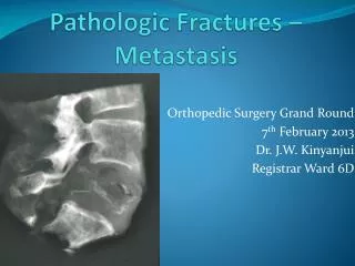
Pediatric Pathologic Fractures Zeke J. Walton, M.D. Lee R. Leddy, M.D. October 2014 Medical University of South Carolina
Pediatric Pathologic Fractures • Objectives • Discuss the principles of management of pediatric pathologic fractures • Define the incidence and demographics of pathologic fractures in children • Describe the most common types of tumor resulting in pathologic fracture and the features of each • Understand the appropriate treatment for each of the most common tumors in pathologic fracture
Pediatric Pathologic Fractures • Surgeon should suspect pathologic fracture when minor trauma results in an injury that is out of proportion to the trauma
Pediatric Pathologic Fractures • A detailed history and physical can be useful in aiding in diagnosis • Plain radiography is usually the first test of choice • Can help define size, location, matrix pattern, growth pattern and periosteal reaction
Pediatric Pathologic Fractures • Differential diagnosis of lesion can be vast but the age of the patient can help narrow the differential • Advanced imaging studies (CT, MRI, Bone scan) are not always required but sometimes can be useful to identify and delineate full extent of the lesion and aid in preoperative planning
Pediatric Pathologic Fractures • Some diagnoses do not require surgery rather the management can be dictated by fracture pattern and typical management for the fracture pattern in absence of tumor • If there is any concern of malignant lesion referral to an orthopaedic oncologist should be made immediately prior to any advanced imaging studies or biopsy
Biopsy • Most lesions will require biopsy at some point, some for diagnostic purposes alone and some for diagnostic and therapeutic purposes • Some benign lesions can be treated by general or pediatric orthopaedic surgeons • Aggressive or malignant appearing lesions should be expeditiously referred to an orthopaedic oncologist prior to biopsy
Biopsy • Biopsy should be made in a location that would not interfere with a future radical resection of a malignant lesion i.e. in line with extensile approach to bone • Minimal soft tissue dissection should be performed during biopsy, rather one should proceed directly down to lesion in line with skin incision • If there is no definitive diagnosis after frozen section and there is any question about the diagnosis one should obtain hemostasis and close in layers ending with subcuticular sutures to await final pathologic diagnosis
Most Common by Age • 0-5 years of age • Benign • Osteomyelitis • Eosinophilic Granuloma • Malignant • Metastatic Tumors (neuroblastoma, Wilm’s) • Leukemia • Ewing’s sarcoma • Fibrosarcoma
Most Common by Age • 5-10 years of age • Benign • Unicameral Bone Cyst (UBC) • Aneurysmal Bone Cyst (ABC) • Non-Ossifying Fibroma (NOF) • Fibrous Dysplasia (FD) • Enchondroma • Osteocondroma • Neurofibroma • Malignant • Leukemia • Osteogenic Sarcoma • Ewing’s Sarcoma
Most Common by Age • 10-20 years of age • Benign • Unicameral Bone Cyst (UBC) • Aneurysmal Bone Cyst (ABC) • Non-Ossifying Fibroma (NOF) • Fibrous Dysplasia (FD) • Enchondroma • Osteocondroma • Chondroblastoma • Giant Cell Tumor • Osteoid Osteoma • Malignant • Leukemia • Lymphoma • Osteogenic Sarcoma • Ewing’s Sarcoma
Unicameral Bone Cyst (UBC) • Benign, expansile fluid-filled lesions most commonly found in the proximal femur and proximal humerus adjacent to physis • Males:females 2.5:1 • 85% under 20 years of age • Approximately 75% of patients will present with a pathologic fracture
Unicameral Bone Cyst • Radiographs typically reveal a well-circumscribed lesion adjacent to physis, usually eccentric in nature. More advanced lesions can show varying degrees of cortical thinning. The fallen-leaf sign is characteristic for UBC, where a fragment of the cyst wall falls to the bottom of the cavity. Differential includes fibrous dysplasia and Aneurismal Bone Cyst (ABC) Pic
Unicameral Bone Cyst • Treatment typically involves treating the fracture first, then the lesion • Upper extremity fractures typically can be treated with immobilization whereas weight bearing bones may require stabilization • Majority of fractures will heal but UBC will persist 20-50% of the time
Unicameral Bone Cyst • UBCs that persist after fracture- goals are to eradicate lesion to prevent future fracture or deformity • There is a current trend towards percutaneous treatments with bone graft substitutes and/or calcium sulfate cement although there is no convincing level 1 evidence to support any treatment method
Aneurysmal Bone Cyst (ABC) • Benign, aggressive lesions most commonly found in femur, humerus, tibia and fibula, skull and posterior elements of the spine • Males:females approximately 1.5:1 • Greatest prevalence around 12 years old • 66% occur in those under 20 years old • 36% of patients will present with a pathologic fracture
Aneurysmal Bone Cyst • Radiographs typically reveal an eccentric, lytic lesion with aggressive features most commonly in the metaphysis. They are usually expansile and cortical thinning is common. There can be periosteal elevation. XRAY MRI
Aneurysmal Bone Cyst • MRI typically reveals internal septations and fluid-fluid levels suggestive of layering of blood components in cystic cavities.
Aneurysmal Bone Cyst • Biopsy is required to rule out telangiectatic osteosarcoma which can have similar appearance on radiographs and MRI • Treatment typically involves extended curettage of lesion with or without adjuvant therapy, bone grafting or placement of PMMA and stabilization
Aneurysmal Bone Cyst • Recurrence rates between 20-40% even for those treated with aggressive intralesional curettage.
Fibrous Dysplasia • Benign non-hereditary condition most commonly found in femur, tibia, pelvis and foot. • Can be monostotic or polyostotic • Polyostotic form can be associated with café-au-lait spots and precocious puberty (McCune-Albright syndrome) or cellular myxomas (Mazabraud’s syndrome)
Fibrous Dysplasia • Represents 5-7% of all benign bone tumors • Patients can present at any age, although there is an increased incidence around 10 years of age • 50% of patients present with pathologic fracture
Fibrous Dysplasia • Radiographs reveal a geographic lesion in the medullary canal usually in the metaphysis or diaphysis. There may be endosteal scalloping and formation of a reactive adjacent cortical thickening. Neither totally lytic or blastic in appearance, it is characterized by ground glass appearance.
Fibrous Dysplasia • Non-displaced pathologic fractures can typically be treated conservatively. • For those that require surgery, curettage with cortical strut graft placement is recommended for slower incorporation and structural support during fracture healing. • Fibrous dysplasia will persist after fracture healing or curettage and bone grafting • All bone grafts, cortical or cancellous are eventually resorbed
Fibrous Dysplasia • Pathologic fractures in the proximal femur are of special concern • Progressive microfractures and deformity over time can lead to shepherd’s crook deformity • Fixation can be accomplished with compression plates, blade plates, cephalomedullary nails or dynamic hip screw with side plates. • It is important to consider the extent of fibrous dysplasia when considering where the distal fixation in the bone will occur
Nonossifying Fibroma (NOF) • Also known as fibrous cortical defect • Most common benign tumor in children • Some estimate 30-40% of those under the age of 20 years old have NOF, mostly asymptomatic
Nonossifying Fibroma • Radiographically NOF are typically geographic, eccentric, metaphyseal lesions that are lytic in nature with a sclerotic rim and cortical erosion. PIC
Nonossifying Fibroma • Low incidence of pathologic fracture • Although when lesions are in the lower extremity, greater than 50% of the diameter of the bone or > 33 mm long they have increased incidence of pathologic fracture • There is evidence that lesions of this size and larger still have less than a 50% risk of fracture
Nonossifying fibroma • Treatment of pathologic fractures similar to UBC, treat fracture first and lesion after • Most can be treated with closed reduction and immobilization
Nonossifying Fibroma • Those who require surgery should undergo curettage and bone grafting with stabilization • Fractures in NOF are known to have the best outcome of all pathologic fractures
Malignant Bone Tumors • Most common primary malignant bone tumors of childhood are osteosarcoma and Ewing’s sarcoma • Pathologic fractures are rare, occurring in 5-15% of osteosarcoma patients and approximately 10% of Ewing’s sarcoma patients • Pathologic fractures are more common in larger, diaphyseal, lytic, fibroblastic or telangiectatic tumors
Malignant Bone Tumors • Standard management of malignant bone tumors typically involves combination of chemotherapy, radiation, and/or surgery after all of the appropriate staging studies have been completed • Initial fracture management include immobilization with sling, splint, cast, traction or external fixation • Careful planning of placement of traction or external fixation pins is important to prevent contamination of soft tissues or margins
Malignant Bone Tumors • Timely referral to a orthopaedic oncologist after conservative stabilization of fracture is important to prevent any delay in treatment • Biopsy and staging studies should be completed by the treating orthopaedic oncologist
Summary • Pathologic fractures can occur from both benign and malignant processes in the pediatric population • Benign conditions can typically be treated conservatively or at the time of surgery • Malignant tumors require a multidisciplinary team at institutions accustomed to treating such patients