Download
1 / 48
480 likes | 597 Vues
Lymphatic System Lecture 12 Bio – 5 JPHubbard. Lymphatic System Organs, vessels and lymph Functions: Produce/maintain lymphocytes – immune response Drains lymph: interstitial fluid & plasma proteins from tissue spaces Transport of substances

E N D
Lymphatic System Lecture 12 Bio – 5 JPHubbard
Lymphatic System • Organs, vessels and lymph • Functions: • Produce/maintain lymphocytes – immune response • Drains lymph: interstitial fluid & plasma proteins from tissue spaces • Transport of substances • dietary lipids & vitamins from GI tract to the blood
What is lymph and where does it come from? • Interstitial Fluid (or tissue fluid) which has entered the lymphatic system • substances being transported from tissues/absorbed from glands and GI tract • chyle (fluid from the intestines after digestion that contains proteins and fats) • dissolved substances: nutrients, wastes, hormones • Cells : • some red blood cells • many white blood cells • Pathogens: viruses and bacteria • Antigens: ‘antibody generating’ – proteins (usually) to which immune system reacts
What is lymph and where does it come from? • What is the function of lymph nodes? • What are the two major duct collecting lymph? • Where do they collect lymph from – and where do they carry it to? • What is the difference between a lymph node and a lymphatic tissue such as the tonsils?
Interstitial Fluid (or tissue fluid) Derived from plasma Hydrostatic pressure (pumping action of heart) forces fluid out of capillaries at arterial end Osmotic pressure: due to concentration of solutes drives return at venous end of capillary bed About 85% of the filtered fluid is returned to the capillary Balance returned via lymphatic vessels (3 liters/day) Edema: swelling – if due to accumulation of excess lymph - lymphedema
Flow of Lymph • Tissue fluid lymph • Flow towards heart promoted by respiratory and muscular movements • Exposure to immune cells takes place in lymph nodes and nodules • Fluid empties into subclavian veins
Lymphatic Capillaries • Blind-ended network through most tissues • Specialized to encourage entry of tissue fluid • Larger in diameter than capillaries • Lack basement lamina • Endothelial cells overlap – provide valve action • Where found/not found • Vascular tissues + • Avascular -
Lymph Circulation • Lymphatic capillaries Lymphatic vessels Lymph nodes Lymphatic ductsLeft and right subclavian veins. • There are five principal trunks. • Lumbar • Intestinal • Subclavian • Jugular • Bronchomediastinal • The five principal trunks pass lymph into 2 main channels= Thoracic duct, Right lymphatic duct.
Vessels unite to form trunks & thoracic ducts • Right side head, arm & chest empty into right lymphatic duct • Rest of body empties into thoracic duct • Lymph is dumped directly into left & right subclavian veins
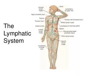
Right jugular trunk Left jugular trunk Right lymphatic duct Left subclavian trunk Left Bronchomediastinal trunk Right subclavian trunk Thoracic duct Left Bronchomediastinal trunk Left lumbar trunk Cisterna chyli Right lumbar trunk Right subclavian vein Left subclavian vein Intestinal trunk
Movement of Lymph: • Similar to movement in veins • Valves encourage one-way flow • Close together – swellings give beaded appearance to vessels • Contractions of smooth muscle in walls of large vessels
Lymphedema: swelling and consequent distention of tissue to blockage of lymphatic vessel • Causes: • Primary: • Absence of/abnormalities of lymphatic tissue • Milroy Disease mutation of FLT4 • Secondary – damage due to: • Disease • Malignancy, • Post-radiation fibrosis
Lymphatic Cells and Lymphatic Organs • Lymphatic or lymphoid cells • Primary lymphatic organs • Site of development of lymphocytes • Secondary lymphatic organs • Site of interaction of lymphocytes with foreign antigen
Lymphatic or Lymphoid Cells • Lymphocytes • Natural Killer Cells • B-cells • T-cells • Macrophages • Others – dendritic cells, reticular cells
Phagocytes (neutrophils & macrophages) • ingest microbes or particulate matter • Fixed and wandering macrophages in specific tissues • histiocytes in the skin, kupffer cells in the liver, alveolar macrophages in the lungs, microglia in the brain & macrophages in spleen, red marrow & lymph nodes • Activate specific immune response • Act as antigen presenting cells stimulate helper T-cells
Natural Killer Cells • NK cells kill a variety of microbes & tumor cells • found in blood, spleen, lymph nodes & red marrow • attack cells displaying abnormal MHC antigens (wrong ‘flag’)
T cell matures in Thymus • Cell-mediated response, ie.- cells that attack • Cytotoxic or Killer T-cells attack non-self cells • bacteria, fungi, viruses, parasites, cancer, and tissue transplants • helper cells (regulatory T-cells) stimulate production of both T and B cell clones • Memory T-Cells: • Reactivate cell line if antigen again appears
B-cells Mature in Bone Marrow • Mature as Plasma cells • Produce antibodies or immunoglobulins • bind to foreign antigen as toxin produced by pathogen • either free or on cell surface - binding may destroy antigen directly, make it a better target for phagocytes • Memory B cells • Reactivated is same antigen again appears
Lymphatic Organs and Lymphocytes Primary lymphatic organs • provide environment for stem cells to divide & mature into B and T lymphocytes • red bone marrow gives rise to mature B cells • thymus is site where pre-T cells from red marrow mature • Secondary lymphatic organs & tissues • site where most immune responses occur • lymph nodes, spleen & lymphatic nodules
Lymphoid Tissues/Organs • Nodules: • Connective tissue dominated by lymphocytes • Lack capsule, generally small (~ 1 mm) • Immune cells act to intercept pathogens crossing epithelium • Lymph Nodes • Separation from surrounding tissue – fibrous connective tissue capsule • Pattern of lymph circulation
Major Lymphoid Tissues - Nodules • MALT – mucosa associated lymphatic tissue • Walls of digestive tract • Peyer’s patches • Walls of appendix • Tonsils – ring throat • single pharyngeal (adenoids) • Paired palatine • Paired lingual
Peyer’s Patches • Ilium of Sm Intestine
Lymph Nodes • ~ oval, to 1 inch • Fibrous connective covering = capsule • Fibrous partitions • Hilus: point of entry of blood vessels, exit of efferent lymphatic vessel • Afferent vessels enter through capsule opposite hilus
Function of Lymph Nodes • Expose lymph to populations of immune cells – ‘filter lymph’ • Exposure to populations of B and T cells • Activate immune system • Due to exposure to dentritic cells – act as antigen presenting cells
Circulation of Lymph Through Lymph Nodes • Subcapsular sinus • Dendritic cells collect antigens, activate immune system • Outercapsule • Aggregates of B-cells • Deep cortex • T-cells • Medulla • Medullary cords – elongate masses of B cells, plasma cells (secrete antibodies) • Exit via Hilus
Distribution of Lymph Nodes • Cervical – head/neck • Axillary – upper limbs, mammary in F. • Supratrochlear – lower arms • Popliteal/Inguinal - from lower limbs • Abdominal – Urinary and reproductive systems • Intestinal/mesenterial lymph nodes + Peyer’s patches from digestive tract • Thoracic – lungs, resp. and mediastinal strs.
Parotid lymph node Preauricular node Facial lymph nodes Retroauricular node Occipital lymph node Submental lymph node Superficial cervical lymph node Submandibular lymph node Deep cervical lymph node
Deltopectoral lymph node Supratrochlear lymph node Axillary
Sternal lymph node Axillary lymph nodes
Superficial inguinal lymph nodes
Lumbar lymph node Common iliac lymph nodes Sacral lymph node Internal iliac lymph node External iliac lymph node
Superior mesenteric nodes Inferior mesenteric lymph node
Anterior mediastinal lymph node Tracheobronchial lymph node Intercostal lymph node Posterior mediastinal lymph node Phrenic lymph node
Thymus Gland • Large organ in infants (70 g) but atrophied as adult (3 g) • 2 lobed organ located in mediastinum • Capsule & trabeculae divideit into lobules • Each lobule has cortex &medulla • Cortex • tightly packed lymphocytes ¯ophages • Medulla • reticular epithelial cells produces thymic hormones • Hassall’s corpuscles
Spleen • Largest lymphoid organ in body • Functions: • Destruction of old erythrocytes • Exposure of blood to populations of immune cells • storage of platelets
Spleen • 5 inch organ between stomach & diaphragm • Hilus contains blood & lymphatic vessels • Stroma consists of capsule, trabeculae, fibers & fibroblasts • Parenchyma consists of white pulp and red pulp • white is lymphatic tissue (lymphocytes & macrophages) around branches of splenic artery • red pulp is venous sinuses filled with blood & splenic tissue (splenic cords)
Thymus • Processes T-lymphocytes or T-cells for function against specific pathogens • Active primarily during childhood and decreases in size with age