Download
1 / 74
940 likes | 1.49k Vues
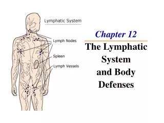
The Lymphatic System and Body Defenses. The Lymphatic System. Consists of two semi-independent parts Lymphatic vessels Lymphoid tissues and organs Lymphatic system functions Transport fluids back to the blood Play essential roles in body defense and resistance to disease.

E N D
The Lymphatic System • Consists of two semi-independent parts • Lymphatic vessels • Lymphoid tissues and organs • Lymphatic system functions • Transport fluids back to the blood • Play essential roles in body defense and resistance to disease
Lymphatic Characteristics • Lymph – excess tissue fluid carried by lymphatic vessels • Properties of lymphatic vessels • One way system toward the heart • No pump • Lymph moves toward the heart • Milking action of skeletal muscle • Rhythmic contraction of smooth muscle in vessel walls
Lymphatic Vessels • Lymph Capillaries • Walls overlap to form flap-like minivalves • Fluid leaks into lymph capillaries • Capillaries are anchored to connective tissue by filaments • Higher pressure on the inside closes minivalves
Lymphatic Vessels Figure 12.2
Lymphatic Vessels • Lymphatic collecting vessels • Collects lymph from lymph capillaries • Carries lymph to and away from lymph nodes Figure 12.1
Lymphatic Vessels • Lymphatic collecting vessels (continued) • Returns fluid to circulatory veins near the heart • Right lymphatic duct • Thoracic duct Figure 12.1
Lymph • Materials returned to the blood • Water • Blood cells • Proteins
Lymph • Harmful materials that enter lymph vessels • Bacteria • Viruses • Cancer cells • Cell debris
Lymph Nodes • Filter lymph before it is returned to the blood • Defense cells within lymph nodes • Macrophages – engulf and destroy foreign substances • Lymphocytes – provide immune response to antigens
Lymph Nodes Figure 12.3
Lymph Node Structure • Most are kidney-shaped, less than 1 inch long • Cortex • Outer part • Contains follicles – collections of lymphocytes • Medulla • Inner part • Contains phagocytic macrophages
Lymph Node Structure Figure 12.4
Flow of Lymph Through Nodes • Lymph enters the convex side through afferent lymphatic vessels • Lymph flows through a number of sinuses inside the node • Lymph exits through efferent lymphatic vessels • Fewer efferent than afferent vessels causes flow to be slowed
Other Lymphoid Organs • Several other organs contribute to lymphatic function • Spleen • Thymus • Tonsils • Peyer’s patches Figure 12.5
The Spleen • Located on the left side of the abdomen • Filters blood • Destroys worn out blood cells • Forms blood cells in the fetus • Acts as a blood reservoir
The Thymus • Located low in the throat, overlying the heart • Functions at peak levels only during childhood • Produces hormones (like thymosin) to program lymphocytes
Tonsils • Small masses of lymphoid tissue around the pharynx • Trap and remove bacteria and other foreign materials • Tonsillitis is caused by congestion with bacteria
Peyer’s Patches • Found in the wall of the small intestine • Resemble tonsils in structure • Capture and destroy bacteria in the intestine
Mucosa-Associated Lymphatic Tissue (MALT) • Includes: • Peyer’s patches • Tonsils • Other small accumulations of lymphoid tissue • Acts as a sentinal to protect respiratory and digestive tracts
Body Defenses • The body is constantly in contact with bacteria, fungi, and viruses • The body has two defense systems for foreign materials • Nonspecific defense system • Specific defense system
Body Defenses • Nonspecific defense system • Mechanisms protect against a variety of invaders • Responds immediately to protect body from foreign materials • Specific defense system • Specific defense is required for each type of invader • Also known as the immune system
Body Defenses Figure 12.6
Nonspecific Body Defenses • Body surface coverings • Intact skin • Mucous membranes • Specialized human cells • Chemicals produced by the body
Surface Membrane Barriers – First Line of Defense • The skin • Physical barrier to foreign materials • pH of the skin is acidic to inhibit bacterial growth • Sebum is toxic to bacteria • Vaginal secretions are very acidic
Surface Membrane Barriers – First Line of Defense • Stomach mucosa • Secretes hydrochloric acid • Has protein-digesting enzymes • Saliva and lacrimal fluid contain lysozyme • Mucus traps microogranisms in digestive and respiratory pathways
Defensive Cells • Phagocytes (neutrophils and macrophages) • Engulfs foreign material into a vacuole • Enzymes from lysosomes digest the material Figure 12.7a
Events of Phagocytosis Figure 12.7b
Defensive Cells • Natural killer cells • Can lyse and kill cancer cells • Can destroy virus- infected cells
Inflammatory Response - Second Line of Defense • Triggered when body tissues are injured • Produces four cardinal signs • Redness • Heat • Swelling • Pain • Results in a chain of events leading to protection and healing
Functions of the Inflammatory Response • Prevents spread of damaging agents • Disposes of cell debris and pathogens • Sets the stage for repair
Steps in the Inflammatory Response Figure 12.8
Antimicrobial Chemicals • Complement • A group of at least 20 plasma proteins • Activated when they encounter and attach to cells (complement fixation) Figure 12.10
Antimicrobial Chemicals • Complement (continued) • Damage foreign cell surfaces • Has vasodilators, chemotaxis, and opsonization Figure 12.10
Antimicrobial Chemicals • Interferon • Secreted proteins of virus-infected cells • Bind to healthy cell surfaces to inhibit viruses binding
Fever • Abnormally high body temperature • Hypothalmus heat regulation can be reset by pyrogens (secreted by white blood cells) • High temperatures inhibit the release of iron and zinc from liver and spleen needed by bacteria • Fever also increases the speed of tissue repair
Specific Defense: The Immune System – Third Line of Defense • Antigen specific – recognizes and acts against particular foreign substances • Systemic – not restricted to the initial infection site • Has memory – recognizes and mounts a stronger attack on previously encountered pathogens
Types of Immunity • Humoral immunity • Antibody-mediated immunity • Cells produce chemicals for defense • Cellular immunity • Cell-mediated immunity • Cells target virus infected cells
Antigens (Nonself) • Any substance capable of exciting the immune system and provoking an immune response • Examples of common antigens • Foreign proteins • Nucleic acids • Large carbohydrates • Some lipids • Pollen grains • Microorganisms
Self-Antigens • Human cells have many surface proteins • Our immune cells do not attack our own proteins • Our cells in another person’s body can trigger an immune response because they are foreign • Restricts donors for transplants
Allergies • Many small molecules (called haptens or incomplete antigens) are not antigenic, but link up with our own proteins • The immune system may recognize and respond to a protein-hapten combination • The immune response is harmful rather than protective because it attacks our own cells
Cells of the Immune System • Lymphocytes • Originate from hemocytoblasts in the red bone marrow • B lymphocytes become immunocompetent in the bone marrow • T lymphocytes become immunocompetent in the thymus • Macrophages • Arise from monocytes • Become widely distributed in lymphoid organs
Activation of Lymphocytes Figure 12.11
Humoral (Antibody-Mediated) Immune Response • B lymphocytes with specific receptors bind to a specific antigen • The binding event activates the lymphocyte to undergo clonal selection • A large number of clones are produced (primary humoral response) PRESS TO PLAY HUMORAL IMMUNITY ANIMATION
Humoral (Antibody Mediated) Immune Response • Most B cells become plasma cells • Produce antibodies to destroy antigens • Activity lasts for four or five days • Some B cells become long-lived memory cells (secondary humoral response)
Humoral Immune Response Figure 12.12
Secondary Response • Memory cells are long-lived • A second exposure causes a rapid response • The secondary response is stronger and longer lasting Figure 12.13
Active Immunity • Your B cells encounter antigens and produce antibodies • Active immunity can be naturally or artificially acquired Figure 12.14
Passive Immunity • Antibodies are obtained from someone else • Conferred naturally from a mother to her fetus • Conferred artificially from immune serum or gamma globulin • Immunological memory does not occur • Protection provided by “borrowed antibodies”