Lymphatic System: Functions
Lymphatic System: Functions. Returns interstitial fluid and leaked plasma proteins back to blood Together with lymphoid organs provide the structural basis of immune system. Venous system. Arterial system. Heart. Lymphatic system:. Lymph duct. Lymph trunk. Lymph node. Lymphatic

Lymphatic System: Functions
E N D
Presentation Transcript
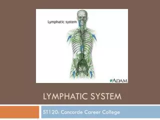
Lymphatic System: Functions • Returns interstitial fluid and leaked plasma proteins back to blood • Together with lymphoid organs provide the structural basis of immune system
Venous system Arterial system Heart Lymphatic system: Lymph duct Lymph trunk Lymph node Lymphatic collecting vessels, with valves Tissue fluid Blood capillaries Lymphatic capillary Tissue cell Blood capillaries Lymphatic capillaries (a) Structural relationship between a capillary bed of the blood vascular system and lymphatic capillaries. Filaments anchored to connective tissue Endothelial cell Flaplike minivalve Fibroblast in loose connective tissue (b) Lymphatic capillaries are blind-ended tubes in which adjacent endothelial cells overlap each other, forming flaplike minivalves. Figure 20.1
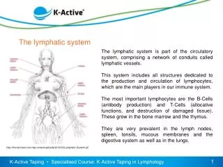
Regional lymph nodes: Internal jugular vein Cervical nodes Entrance of right lymphatic duct into vein Entrance of thoracic duct into vein Axillary nodes Thoracic duct Cisterna chyli Aorta Inguinal nodes Lymphatic collecting vessels Drained by the right lymphatic duct Drained by the thoracic duct (a) General distribution of lymphatic collecting vessels and regional lymph nodes. Figure 20.2a
Lymphoid Cells • Lymphocytes -warriors of immune system • Two types • T cells (T lymphocytes) • B cells (B lymphocytes)
Lymphocytes • T cells and B cells protect against antigens • Anything the body perceives as foreign • Bacteria and their toxins; viruses • Mismatched RBCs or cancer cells • Provides a proliferation site for lymphocytes • Furnishes a surveillance vantage point
Macrophage Reticular cells on reticular fibers Lymphocytes Medullary sinus Reticular fiber Figure 20.3
Afferent lymphatic vessels Cortex • Lymphoid follicle • Germinal center • Subcapsular sinus Efferent lymphatic vessels Follicles Trabecula Subcapsular sinus Capsule Hilum Medulla: • Medullary cord • Medullary sinus Medullary cords Medullary sinuses Trabeculae Capsule (b) Photomicrograph of part of a lymph node (72x) (a) Longitudinal view of the internal structure of a lymph node and associated lymphatics Figure 20.4
Spleen • Largest lymphoid organ • Functions • Site of lymphocyte proliferation and immune surveillance and response • Cleanses the blood of aged cells and platelets and debris
Diaphragm Spleen Adrenal gland Left kidney Splenic artery Pancreas (c) Photograph of the spleen in its normal position in the abdominal cavity, anterior view. Figure 20.6c
Thymus • Size with age • Infants, in inferior neck • Increases in size, most active during childhood • Stops growing during adolescence and then gradually atrophies • It functions strictly in T lymphocyte maturation
Thymic (Hassall’s) corpuscle Medulla Cortex Figure 20.7
Pharyngeal tonsil Palatine tonsil Lingual tonsil Tonsil Tonsillar crypt Germinal centers in lymphoid follicles Figure 20.8
Immunity • Resistance to disease • Immune system has two parts • Innate (nonspecific) defense system • Adaptive (specific) defense system
Immunity • Innate defense system has two lines of defense • First line of defense is external body membranes (skin and mucosae) • Second line of defense is antimicrobial proteins, phagocytes, and other cells • Inhibit spread of invaders • Inflammation is its most important mechanism
Immunity • Adaptive defense system • Third line of defense attacks particular foreign substances • Takes longer to react than the innate system • Innate and adaptive defenses are deeply intertwined
Surface barriers • Skin • Mucous membranes Innate defenses Internal defenses • Phagocytes • NK cells • Inflammation • Antimicrobial proteins • Fever Humoral immunity • B cells Adaptive defenses Cellular immunity • T cells Figure 21.1
1 Phagocyte adheres to pathogens or debris. 2 Phagocyte forms pseudopods that eventually engulf the particles forming a phagosome. Phagosome (phagocytic vesicle) Lysosome 3 Lysosome fuses with the phagocytic vesicle, forming a phagolysosome. Acid hydrolase enzymes 4 Lysosomal enzymes digest the particles, leaving a residual body. 5 Exocytosis of the vesicle removes indigestible and residual material. (b) Events of phagocytosis. Figure 21.2b
Inflammatory Response • Cardinal signs of acute inflammation: • Redness • Heat • Swelling • Pain (And sometimes 5. Impairment of function)
Innate defenses Internal defenses Tissue injury Release of chemical mediators (histamine, complement, kinins, prostaglandins, etc.) Release of leukocytosis- inducing factor Leukocytosis (increased numbers of white blood cells in bloodstream) Vasodilation of arterioles Increased capillary permeability Attract neutrophils, monocytes, and lymphocytes to area (chemotaxis) Leukocytes migrate to injured area Local hyperemia (increased blood flow to area) Capillaries leak fluid (exudate formation) Margination (leukocytes cling to capillary walls) Initial stimulus Physiological response Signs of inflammation Diapedesis (leukocytes pass through capillary walls) Leaked clotting proteins form interstitial clots that wall off area to prevent injury to surrounding tissue Leaked protein-rich fluid in tissue spaces Result Phagocytosis of pathogens and dead tissue cells (by neutrophils, short-term; by macrophages, long-term) Heat Redness Pain Swelling Temporary fibrin patch forms scaffolding for repair Locally increased temperature increases metabolic rate of cells Possible temporary limitation of joint movement Pus may form Area cleared of debris Healing Figure 21.3
Phagocyte Mobilization • Neutrophils, then phagocytes flood to inflamed sites
Innatedefenses Internaldefenses Inflammatorychemicalsdiffusingfrom theinflamed siteact as chemotacticagents. 4 Chemotaxis.Neutrophilsfollow chemicaltrail. Capillary wall Basementmembrane Endothelium 1 2 3 Leukocytosis.Neutrophils enter bloodfrom bone marrow. Margination.Neutrophils clingto capillary wall. Diapedesis.Neutrophils flatten andsqueeze out of capillaries. Figure 21.4
Antimicrobial Proteins • Interferons (IFNs) and complement proteins • Attack microorganisms directly • Hinder microorganisms’ ability to reproduce
Alternative pathway Classical pathway Spontaneous activation Antigen-antibody complex + + Stabilizing factors (B, D, and P) + complex No inhibitors on pathogen surface Enhances inflammation: Opsonization: stimulates histamine release, increases blood vessel permeability, attracts phagocytes by chemotaxis, etc. coats pathogen surfaces, which enhances phagocytosis Insertion of MAC and cell lysis (holes in target cell’s membrane) Pore Complement proteins (C5b–C9) Membrane of target cell Figure 21.6
Fever • Systemic response • Leukocytes secrete pyrogens • Pyrogens reset the body’s thermostat upward • High fevers are dangerous because heat denatures enzymes • Benefits of moderate fever • Causes the liver to sequester iron and zinc • Increases metabolic rate, speeds up repair
Adaptive Defenses • Adaptive immune response • Is specific • Is systemic • Has memory • Two separate overlapping arms • Humoral (antibody-mediated) immunity • Cellular (cell-mediated) immunity
Antigens • Substances that can mobilize the adaptive defenses and provoke an immune response • Most are large, complex molecules not normally found in the body (nonself)
Antigen- binding sites Antigenic determinants Antibody A Antigen Antibody B Antibody C Figure 21.7
Self-Antigens • Protein molecules (self-antigens) on the surface of cells • Antigenic to others in transfusions or grafts • Example: MHC proteins • Coded for by genes of the major histocompatibility complex (MHC) and are unique to an individual
Cells of the Adaptive Immune System • Two types of lymphocytes • B lymphocytes (B cells)—humoral immunity • T lymphocytes (T cells)—cell-mediated immunity • Antigen-presenting cells (APCs) • Do not respond to specific antigens
Red bone marrow: site of lymphocyte origin Humoral immunity Adaptive defenses Cellular immunity Primary lymphoid organs: site of development of immunocompetence as B or T cells Immature lymphocytes Red bone marrow Secondary lymphoid organs: site of antigen encounter, and activation to become effector and memory B or T cells 1 Lymphocytes destined to become T cells migrate (in blood) to the thymus and develop immunocompetence there. B cells develop immunocompetence in red bone marrow. Thymus Bone marrow 2 Immunocompetent but still naive lymphocytes leave the thymus and bone marrow. They “seed” the lymph nodes, spleen, and other lymphoid tissues where they encounter their antigen. Lymph nodes, spleen, and other lymphoid tissues 3 Antigen-activated immunocompetent lymphocytes (effector cells and memory cells) circulate continuously in the bloodstream and lymph and throughout the lymphoid organs of the body. Figure 21.8
Clonal Selection • B cell is activated when antigens bind to its surface receptors • Bound antigen enters cell • Stimulated B cell grows to form a clone of identical cells bearing the same antigen-specific receptors(T cells required to help B cells achieve full activation)
Fate of the Clones • Most clone cells become plasma cells • secrete antibodies at rate of 2000 per second for 4 - 5days • Secreted antibodies • Circulate in blood or lymph • Bind to free antigens • Mark the antigens for destruction
Adaptive defenses Humoral immunity Antigen Primary response (initial encounter with antigen) Antigen binding to a receptor on a specific B lymphocyte (B lymphocytes with non-complementary receptors remain inactive) Proliferation to form a clone Activated B cells Plasma cells (effector B cells) Memory B cell— primed to respond to same antigen Secreted antibody molecules Figure 21.11 (1 of 2)
Immunological Memory • Primary immune response • Occurs on first exposure to specific antigen • Lag period: three to six days • Peak levels of antibody reached in 10 days • Antibody levels then decline
Immunological Memory • Secondary immune response • Occurs on re-exposure to same antigen • Sensitized memory cells respond within hours • Antibody levels peak in 2 – 3 d at higher levels • Antibodies bind with greater affinity • Antibody level remain high for weeks to months
Adaptive defenses Humoral immunity Antigen Primary response (initial encounter with antigen) Antigen binding to a receptor on a specific B lymphocyte (B lymphocytes with non-complementary receptors remain inactive) Proliferation to form a clone Activated B cells Plasma cells (effector B cells) Memory B cell— primed to respond to same antigen Secreted antibody molecules Subsequent challenge by same antigen results in more rapid response Secondary response (can be years later) Clone of cells identical to ancestral cells Plasma cells Secreted antibody molecules Memory B cells Figure 21.11
Secondary immune response to antigen A is faster and larger; primary immune response to antigen B is similar to that for antigen A. Primary immune response to antigen A occurs after a delay. Anti- bodies to B Anti- bodies to A Second exposure to antigen A; first exposure to antigen B First exposure to antigen A Time (days) Figure 21.12
Humoral immunity Active Passive Naturally acquired Naturally acquired Artificially acquired Artificially acquired Infection; contact with pathogen Antibodies pass from mother to fetus via placenta; or to infant in her milk Vaccine; dead or attenuated pathogens Injection of immune serum (gamma globulin) Figure 21.13
Antigen-binding site Heavy chain variable region Hinge region Heavy chain constant region Stem region Light chain variable region Light chain constant region Disulfide bond (a) Figure 21.14a
Adaptive defenses Humoral immunity Antigen-antibody complex Antigen Antibody Inactivates by Fixes and activates Neutralization (masks dangerous parts of bacterial exotoxins; viruses) Agglutination (cell-bound antigens) Precipitation (soluble antigens) Complement Enhances Enhances Leads to Inflammation Phagocytosis Cell lysis Chemotaxis Histamine release Figure 21.15
Monoclonal Antibodies • Commercially prepared pure antibody • Proliferate indefinitely and have the ability to produce a single type of antibody • Used in research, clinical testing, and cancer treatment
Cell-Mediated Immune Response • T cells defend against intracellular antigens • Major types of T cells • CD4 cells become helper T cells (TH) • CD8 cells become cytotoxic T cells (TC) that destroy cells harboring foreign antigens • Other types of T cells • Regulatory T cells (TREG) • Memory T cells
Adaptive defenses Cellular immunity Immature lymphocyte Red bone marrow T cell receptor T cell receptor Maturation Class I MHC protein Class II MHC protein CD4 cell CD8 cell Thymus Activation Activation APC (dendritic cell) Memory cells APC (dendritic cell) CD4 CD8 Lymphoid tissues and organs Effector cells Helper T cells (or regulatory T cells) Cytotoxic T cells Blood plasma Figure 21.16
T Cell Activation • APCs migrate to lymph nodes to present their antigens to T cells • T cell activation is a two-step process • Antigen binding • Co-stimulation
Adaptive defenses Cellular immunity Viral antigen 1 Dendritic cell engulfs an exogenous antigen, processes it, and displays its fragments on class II MHC protein. Class lI MHC protein displaying processed viral antigen Dendritic cell CD4 protein 2 Immunocompetent CD4 cell recognizes antigen-MHC complex. Both TCR and CD4 protein bind to antigen-MHC complex. T cell receptor (TCR) Immunocom- petent CD4 T cell Clone formation 3 CD4 cells are activated, proliferate (clone), and become memory and effector cells. Helper T memory cell Activated helper T cells Figure 21.18
Roles of Helper T(TH) Cells • Play a central role in the adaptive immune response • Once primed by APC presentation of antigen, they • Help activate T and B cells • Induce T and B cell proliferation • Activate macrophages and recruit other immune cells • Without TH, there is no immune response
TH cell help in humoral immunity Activated helper T cell TH cell binds with the self-nonself complexes of a B cell that has encountered its antigen and is displaying it on MHC II on its surface. 1 T cell receptor (TCR) Helper T cell CD4 protein MHC II protein of B cell displaying processed antigen TH cell releases interleukins as co-stimulatory signals to complete B cell activation. 2 IL- 4 and other cytokines B cell (being activated) (a) Figure 21.19a
TH cell help in cell-mediated immunity CD4 protein 1 Previously activated TH cell binds dendritic cell. Helper T cell Class II MHC protein APC (dendritic cell) 2 TH cell stimulates dendritic cell to express co-stimulatory molecules (not shown) needed to activate CD8 cell. IL-2 3 Dendritic cell can now activate CD8 cell with the help of interleukin 2 secreted by TH cell. Class I MHC protein CD8 protein CD8 T cell (b) Figure 21.19b
Roles of Cytotoxic T(TC) Cells • Targets • Virus-infected cells • Cells with intracellular bacteria or parasites • Cancer cells • Foreign cells (transfusions or transplants)